- PDB-3n6r: CRYSTAL STRUCTURE OF the holoenzyme of PROPIONYL-COA CARBOXYLASE (PCC) -
+
Open data
ID or keywords:
Loading...
-
Basic information
Entry
Database: PDB / ID: 3n6r
Title
CRYSTAL STRUCTURE OF the holoenzyme of PROPIONYL-COA CARBOXYLASE (PCC)
Components
Propionyl-CoA carboxylase, alpha subunit
Propionyl-CoA carboxylase, beta subunit
Keywords
LIGASE / protein complex / biotin-dependent carboxylase
Function / homology
Function and homology information
propionyl-CoA carboxylase / propionyl-CoA carboxylase activity / lipid catabolic process / ATP binding / metal ion binding Similarity search - Function
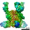
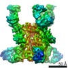
Journal: Nature / Year: 2010 Title: Crystal structure of the alpha(6)beta(6) holoenzyme of propionyl-coenzyme A carboxylase. Authors: Christine S Huang / Kianoush Sadre-Bazzaz / Yang Shen / Binbin Deng / Z Hong Zhou / Liang Tong / Abstract: Propionyl-coenzyme A carboxylase (PCC), a mitochondrial biotin-dependent enzyme, is essential for the catabolism of the amino acids Thr, Val, Ile and Met, cholesterol and fatty acids with an odd ...Propionyl-coenzyme A carboxylase (PCC), a mitochondrial biotin-dependent enzyme, is essential for the catabolism of the amino acids Thr, Val, Ile and Met, cholesterol and fatty acids with an odd number of carbon atoms. Deficiencies in PCC activity in humans are linked to the disease propionic acidaemia, an autosomal recessive disorder that can be fatal in infants. The holoenzyme of PCC is an alpha(6)beta(6) dodecamer, with a molecular mass of 750 kDa. The alpha-subunit contains the biotin carboxylase (BC) and biotin carboxyl carrier protein (BCCP) domains, whereas the beta-subunit supplies the carboxyltransferase (CT) activity. Here we report the crystal structure at 3.2-A resolution of a bacterial PCC alpha(6)beta(6) holoenzyme as well as cryo-electron microscopy (cryo-EM) reconstruction at 15-A resolution demonstrating a similar structure for human PCC. The structure defines the overall architecture of PCC and reveals unexpectedly that the alpha-subunits are arranged as monomers in the holoenzyme, decorating a central beta(6) hexamer. A hitherto unrecognized domain in the alpha-subunit, formed by residues between the BC and BCCP domains, is crucial for interactions with the beta-subunit. We have named it the BT domain. The structure reveals for the first time the relative positions of the BC and CT active sites in the holoenzyme. They are separated by approximately 55 A, indicating that the entire BCCP domain must translocate during catalysis. The BCCP domain is located in the active site of the beta-subunit in the current structure, providing insight for its involvement in the CT reaction. The structural information establishes a molecular basis for understanding the large collection of disease-causing mutations in PCC and is relevant for the holoenzymes of other biotin-dependent carboxylases, including 3-methylcrotonyl-CoA carboxylase (MCC) and eukaryotic acetyl-CoA carboxylase (ACC).
History
Deposition
May 26, 2010
Deposition site: RCSB / Processing site: RCSB
Revision 1.0
Aug 25, 2010
Provider: repository / Type: Initial release
Revision 1.1
Jul 13, 2011
Group: Source and taxonomy / Version format compliance
Monochromator: SI(111) / Protocol: SINGLE WAVELENGTH / Monochromatic (M) / Laue (L): M / Scattering type: x-ray
Radiation wavelength
Wavelength: 1.0809 Å / Relative weight: 1
Reflection
Resolution: 3.2→30 Å / Num. obs: 169648 / % possible obs: 92 % / Redundancy: 1.9 % / Rmerge(I) obs: 0.084 / Net I/σ(I): 8.6854
Reflection shell
Resolution: 3.2→3.31 Å / Redundancy: 1.7 % / Rmerge(I) obs: 0.342 / Mean I/σ(I) obs: 2.024 / % possible all: 80
-
Processing
Software
Name
Version
Classification
CBASS
datacollection
PHASER
phasing
CNS
1.2
refinement
DENZO
datareduction
SCALEPACK
datascaling
Refinement
Method to determine structure: MOLECULAR REPLACEMENT / Resolution: 3.2→29.36 Å / Rfactor Rfree error: 0.002 / Data cutoff high absF: 194743.4 / Data cutoff low absF: 0 / Isotropic thermal model: RESTRAINED / Cross valid method: THROUGHOUT / σ(F): 0 Details: BULK SOLVENT MODEL USED. THE RESIDUES ARE NUMBERED ACCORDING TO THE HUMAN PCC SEQUENCE
In the structure databanks used in Yorodumi, some data are registered as the other names, "COVID-19 virus" and "2019-nCoV". Here are the details of the virus and the list of structure data.
Jan 31, 2019. EMDB accession codes are about to change! (news from PDBe EMDB page)
EMDB accession codes are about to change! (news from PDBe EMDB page)
The allocation of 4 digits for EMDB accession codes will soon come to an end. Whilst these codes will remain in use, new EMDB accession codes will include an additional digit and will expand incrementally as the available range of codes is exhausted. The current 4-digit format prefixed with “EMD-” (i.e. EMD-XXXX) will advance to a 5-digit format (i.e. EMD-XXXXX), and so on. It is currently estimated that the 4-digit codes will be depleted around Spring 2019, at which point the 5-digit format will come into force.
The EM Navigator/Yorodumi systems omit the EMD- prefix.
Related info.:Q: What is EMD? / ID/Accession-code notation in Yorodumi/EM Navigator
Yorodumi is a browser for structure data from EMDB, PDB, SASBDB, etc.
This page is also the successor to EM Navigator detail page, and also detail information page/front-end page for Omokage search.
The word "yorodu" (or yorozu) is an old Japanese word meaning "ten thousand". "mi" (miru) is to see.
Related info.:EMDB / PDB / SASBDB / Comparison of 3 databanks / Yorodumi Search / Aug 31, 2016. New EM Navigator & Yorodumi / Yorodumi Papers / Jmol/JSmol / Function and homology information / Changes in new EM Navigator and Yorodumi
 Movie
Movie Controller

 Yorodumi
 Open data
 Basic information
 Components
 Keywords
 Function and homology information
 Ruegeria pomeroyi (bacteria)
 X-RAY DIFFRACTION /
  Authors
 Citation
 Structure visualization
 Downloads & links
 Other downloads

 PDBj
PDBj


 Assembly

 Mass: 228.311 Da / Num. of mol.: 6 / Source method: obtained synthetically / Formula: C10H16N2O2S
 Sample preparation
 Processing