- PDB-3n5l: Crystal structure of a binding protein component of ABC phosphona... -
+
Open data
ID or keywords:
Loading...
-
Basic information
Entry
Database: PDB / ID: 3n5l
Title
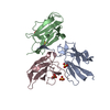
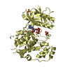
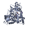
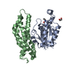
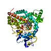
Crystal structure of a binding protein component of ABC phosphonate transporter (PA3383) from Pseudomonas aeruginosa at 1.97 A resolution
Components
Binding protein component of ABC phosphonate transporter
Keywords
TRANSPORT PROTEIN / Structural Genomics / Joint Center for Structural Genomics / JCSG / Protein Structure Initiative / PSI-2 / ABC transport system / periplasmic phosphonate-binding
Function / homology
Function and homology information
organic phosphonate transport / cell envelope / ATP-binding cassette (ABC) transporter complex / transmembrane transport Similarity search - Function
Mass: 18.015 Da / Num. of mol.: 636 / Source method: isolated from a natural source / Formula: H2O
-
Details
Has protein modification
Y
Sequence details
(1) THE CONSTRUCT (RESIDUES 26-334) WAS EXPRESSED WITH AN N-TERMINAL PURIFICATION TAG ...(1) THE CONSTRUCT (RESIDUES 26-334) WAS EXPRESSED WITH AN N-TERMINAL PURIFICATION TAG MGSDKIHHHHHHENLYFQG. THE TAG WAS REMOVED WITH TEV PROTEASE LEAVING ONLY A GLYCINE (0) FOLLOWED BY THE TARGET SEQUENCE. (2) DNA SEQUENCING OF THE CLONED CONSTRUCT SHOWS A MIXTURE OF BOTH "CCC" (PROLINE) AND "GCC" (ALANINE) AT THE POSITION CORRESPONDING TO RESIDUE 272. THE REST OF THE SEQUENCE TRACE IS CLEAN. A MIXTURE OF PROLINE AND ALANINE AT POSITION 272 IS CONSISTENT WITH THE MASS SPECTROMETRY PROFILE OF THE PURIFIED PROTEIN. HOWEVER, THE CRYSTAL APPEARS TO CONTAIN PREDOMINANTLY ALANINE AT POSITION 272 BASED ON THE ELECTRON DENSITY FIT AND CRYSTAL PACKING CONSTRAINTS. MICROHETEROGENEITY PRESENT AT POSITION 272 WAS NOT MODELED.
-
Experimental details
-
Experiment
Experiment
Method: X-RAY DIFFRACTION / Number of used crystals: 1
-
Sample preparation
Crystal
Density Matthews: 3.46 Å3/Da / Density % sol: 64.42 %
Crystal grow
Temperature: 277 K / Method: vapor diffusion, sitting drop / pH: 7 Details: 2.000000000M (NH4)2SO4, 0.200000000M Li2SO4, 0.1M TRIS pH 7.0, NANODROP, VAPOR DIFFUSION, SITTING DROP, temperature 277K
Resolution: 1.97→49.445 Å / Num. obs: 68965 / % possible obs: 100 % / Observed criterion σ(I): -3 / Biso Wilson estimate: 25.138 Å2 / Rmerge(I) obs: 0.131 / Net I/σ(I): 12.37
Reflection shell
Resolution (Å)
Rmerge(I) obs
Mean I/σ(I) obs
Num. measured obs
Num. unique obs
Diffraction-ID
% possible all
1.97-2.05
0.966
2.1
56754
7646
1
100
2.05-2.12
0.725
2.8
42950
5795
1
100
2.12-2.21
0.562
3.7
47865
6421
1
100
2.21-2.31
0.443
4.7
44565
6028
1
100
2.31-2.43
0.346
5.9
44468
5961
1
100
2.43-2.58
0.291
7.1
44831
6018
1
100
2.58-2.78
0.224
9.1
45955
6185
1
100
2.78-3.06
0.149
13.1
45595
6130
1
100
3.06-3.5
0.086
20.8
45397
6132
1
100
3.5-4.4
0.051
31.9
45603
6221
1
100
4.4-49.445
0.046
35.2
46085
6513
1
99.8
-
Phasing
Phasing
Method: MAD
-
Processing
Software
Name
Version
Classification
NB
REFMAC
5.5.0110
refinement
PHENIX
refinement
SHELX
phasing
MolProbity
3beta29
modelbuilding
XSCALE
datascaling
PDB_EXTRACT
3.006
dataextraction
XDS
datareduction
SHELXD
phasing
autoSHARP
phasing
Refinement
Method to determine structure: MAD / Resolution: 1.97→49.445 Å / Cor.coef. Fo:Fc: 0.965 / Cor.coef. Fo:Fc free: 0.949 / Occupancy max: 1 / Occupancy min: 0.3 / SU B: 5.02 / SU ML: 0.074 / Cross valid method: THROUGHOUT / σ(F): 0 / ESU R Free: 0.113 Stereochemistry target values: MAXIMUM LIKELIHOOD WITH PHASES Details: 1. HYDROGENS HAVE BEEN ADDED IN THE RIDING POSITIONS. 2. A MET-INHIBITION PROTOCOL WAS USED FOR SELENOMETHIONINE INCORPORATION DURING PROTEIN EXPRESSION. THE OCCUPANCY OF THE SE ATOMS IN THE ...Details: 1. HYDROGENS HAVE BEEN ADDED IN THE RIDING POSITIONS. 2. A MET-INHIBITION PROTOCOL WAS USED FOR SELENOMETHIONINE INCORPORATION DURING PROTEIN EXPRESSION. THE OCCUPANCY OF THE SE ATOMS IN THE MSE RESIDUES WAS REDUCED TO 0.75 FOR THE REDUCED SCATTERING POWER DUE TO PARTIAL S-MET INCORPORATION. 3. ATOM RECORD CONTAINS SUM OF TLS AND RESIDUAL B FACTORS. ANISOU RECORD CONTAINS SUM OF TLS AND RESIDUAL U FACTORS. 4. WATERS WERE EXCLUDED FROM AUTOMATIC TLS ASSIGNMENT. 5. A UNKNOWN LIGAND (UNL), RESEMBLING GLYCEROL-3-PHOSPHATE WAS MODELED INTO THE BINDING SITE OF EACH MONOMER. THE PROTEIN IS A HOMOLOG OF PHND OF E. COLI, WHICH BINDS ALKYLPHOSPHONATE. ETHYLENE GLYCOL (EDO), SULFATE (SO4) AND CHLORIDE (CL) MODELED ARE PRESENT PROTEIN/CRYSTALLIZATION/CRYO BUFFER.
Rfactor
Num. reflection
% reflection
Selection details
Rfree
0.191
3488
5.1 %
RANDOM
Rwork
0.158
-
-
-
obs
0.16
68909
99.89 %
-
Solvent computation
Ion probe radii: 0.8 Å / Shrinkage radii: 0.8 Å / VDW probe radii: 1.2 Å / Solvent model: MASK
In the structure databanks used in Yorodumi, some data are registered as the other names, "COVID-19 virus" and "2019-nCoV". Here are the details of the virus and the list of structure data.
Jan 31, 2019. EMDB accession codes are about to change! (news from PDBe EMDB page)
EMDB accession codes are about to change! (news from PDBe EMDB page)
The allocation of 4 digits for EMDB accession codes will soon come to an end. Whilst these codes will remain in use, new EMDB accession codes will include an additional digit and will expand incrementally as the available range of codes is exhausted. The current 4-digit format prefixed with “EMD-” (i.e. EMD-XXXX) will advance to a 5-digit format (i.e. EMD-XXXXX), and so on. It is currently estimated that the 4-digit codes will be depleted around Spring 2019, at which point the 5-digit format will come into force.
The EM Navigator/Yorodumi systems omit the EMD- prefix.
Related info.:Q: What is EMD? / ID/Accession-code notation in Yorodumi/EM Navigator
Yorodumi is a browser for structure data from EMDB, PDB, SASBDB, etc.
This page is also the successor to EM Navigator detail page, and also detail information page/front-end page for Omokage search.
The word "yorodu" (or yorozu) is an old Japanese word meaning "ten thousand". "mi" (miru) is to see.
Related info.:EMDB / PDB / SASBDB / Comparison of 3 databanks / Yorodumi Search / Aug 31, 2016. New EM Navigator & Yorodumi / Yorodumi Papers / Jmol/JSmol / Function and homology information / Changes in new EM Navigator and Yorodumi
 Movie
Movie Controller

 Yorodumi
 Open data
 Basic information
 Components
 Keywords
 Function and homology information
 Pseudomonas aeruginosa (bacteria)
 X-RAY DIFFRACTION /
  Authors
 Citation
 Structure visualization
 Downloads & links
 Other downloads


 PDBj
PDBj

 Assembly
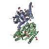


 Mass: 96.063 Da / Num. of mol.: 6 / Source method: obtained synthetically / Formula: SO4
 Mass: 62.068 Da / Num. of mol.: 22 / Source method: obtained synthetically / Formula: C2H6O2
 Mass: 35.453 Da / Num. of mol.: 1 / Source method: obtained synthetically / Formula: Cl
 Sample preparation
 / Beamline: BL9-2 / Wavelength: 0.91837,0.97936
 Processing