 Movie
Movie Controller

[English] 日本語
 Yorodumi
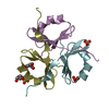
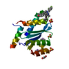
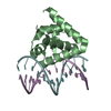
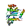
- PDB-3n3f: Crystal Structure of the Human Collagen XV Trimerization Domain: ... -
+ Open data

- Basic information
| Entry | Database: PDB / ID: 3n3f | ||||||
|---|---|---|---|---|---|---|---|
| Title | Crystal Structure of the Human Collagen XV Trimerization Domain: A Potent Trimerizing Unit Common to Multiplexin Collagens | ||||||
 Components Components | Collagen alpha-1(XV) chain | ||||||
 Keywords Keywords | PROTEIN BINDING / collagen / extracellular matrix / basement membrane / collagen XV / trimerization domain / folding / association / chain selection / triple helix / multiplexin / collagen XVIII | ||||||
| Function / homology |  Function and homology information Function and homology informationcollagen type XV trimer / Collagen chain trimerization / extracellular matrix structural constituent conferring tensile strength / Collagen biosynthesis and modifying enzymes / Assembly of collagen fibrils and other multimeric structures / Collagen degradation / basement membrane / : / angiogenesis / cell differentiation ...collagen type XV trimer / Collagen chain trimerization / extracellular matrix structural constituent conferring tensile strength / Collagen biosynthesis and modifying enzymes / Assembly of collagen fibrils and other multimeric structures / Collagen degradation / basement membrane / : / angiogenesis / cell differentiation / cell adhesion / endoplasmic reticulum lumen / endoplasmic reticulum / signal transduction / extracellular space / extracellular exosome / extracellular region / membrane Similarity search - Function | ||||||
| Biological species |  Homo sapiens (human) Homo sapiens (human) | ||||||
| Method |  X-RAY DIFFRACTION / MOLECULAR REPLACEMENT / molecular replacement / Resolution: 2.005 Å X-RAY DIFFRACTION / MOLECULAR REPLACEMENT / molecular replacement / Resolution: 2.005 Å | ||||||
 Authors Authors | Wirz, J.A. | ||||||
 Citation Citation | Journal: Matrix Biol. / Year: 2011 Title: Crystal structure of the human collagen XV trimerization domain: A potent trimerizing unit common to multiplexin collagens. Authors: Wirz, J.A. / Boudko, S.P. / Lerch, T.F. / Chapman, M.S. / Bachinger, H.P. | ||||||
| History |
|
- Structure visualization
| Structure viewer | Molecule: MolmilJmol/JSmol |
|---|
- Downloads & links
-Download
| PDBx/mmCIF format | 3n3f.cif.gz | 34.3 KB | Display | PDBx/mmCIF format |
|---|---|---|---|---|
| PDB format | pdb3n3f.ent.gz | 23.3 KB | Display | PDB format |
| PDBx/mmJSON format | 3n3f.json.gz | Tree view | PDBx/mmJSON format | |
| Others |  Other downloads Other downloads |
-Validation report
| Arichive directory | https://data.pdbj.org/pub/pdb/validation_reports/n3/3n3fftp://data.pdbj.org/pub/pdb/validation_reports/n3/3n3f | HTTPS FTP |
|---|
-Related structure data
| Related structure data | 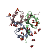 3hshS S: Starting model for refinement |
|---|---|
| Similar structure data |


-Links
 PDBj
PDBj


- Assembly
| Deposited unit | 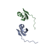
| ||||||||
|---|---|---|---|---|---|---|---|---|---|
| 1 | 
| ||||||||
| 2 | 
| ||||||||
| Unit cell |
|
-Components
| #1: Protein | Mass: 6259.278 Da / Num. of mol.: 2 / Fragment: UNP residues 1133-1186 Source method: isolated from a genetically manipulated source Source: (gene. exp.) Homo sapiens (human) / Gene: COL15A1 / Plasmid: pET23-HisTag-Trx / Production host:  #2: Water | ChemComp-HOH / |  Mass: 18.015 Da / Num. of mol.: 32 / Source method: isolated from a natural source / Formula: H2O Mass: 18.015 Da / Num. of mol.: 32 / Source method: isolated from a natural source / Formula: H2O |
|---|
-Experimental details
-Experiment
| Experiment | Method: X-RAY DIFFRACTION / Number of used crystals: 1 |
|---|
- Sample preparation
| Crystal | Density Matthews: 1.82 Å3/Da / Density % sol: 32.6 % |
|---|---|
| Crystal grow | Temperature: 298 K / Method: vapor diffusion, hanging drop / pH: 7.5 Details: 0.1M Tris, 0.2 M MgCl2, 30% (w/v) PEG3350, pH 7.5, VAPOR DIFFUSION, HANGING DROP, temperature 298K |
-Data collection
| Diffraction | Mean temperature: 100 K |
|---|---|
| Diffraction source | Source: ROTATING ANODE / Type: RIGAKU RU200 / Wavelength: 1.54 Å |
| Detector | Type: RIGAKU RAXIS / Detector: IMAGE PLATE / Date: Nov 19, 2009 |
| Radiation | Monochromator: Ni Filter / Protocol: SINGLE WAVELENGTH / Monochromatic (M) / Laue (L): M / Scattering type: x-ray |
| Radiation wavelength | Wavelength: 1.54 Å / Relative weight: 1 |
| Reflection | Resolution: 2.005→42.539 Å / Num. all: 6095 / Num. obs: 6095 / % possible obs: 100 % / Observed criterion σ(F): 1 / Observed criterion σ(I): 1 / Redundancy: 6.6 % / Biso Wilson estimate: 22.838 Å2 / Rmerge(I) obs: 0.034 / Net I/σ(I): 35.4 |
| Reflection shell | Resolution: 2.005→2.01 Å / Redundancy: 6.5 % / Rmerge(I) obs: 0.11 / Mean I/σ(I) obs: 15.6 / Num. unique all: 881 / % possible all: 100 |
-Phasing
| Phasing | Method: molecular replacement | |||||||||
|---|---|---|---|---|---|---|---|---|---|---|
| Phasing MR | Rfactor: 0.49 / Cor.coef. Fo:Fc: 0.48
|
- Processing
| Software |
| ||||||||||||||||||||||||||||
|---|---|---|---|---|---|---|---|---|---|---|---|---|---|---|---|---|---|---|---|---|---|---|---|---|---|---|---|---|---|
| Refinement | Method to determine structure: MOLECULAR REPLACEMENT Starting model: PDB Entry 3HSH Resolution: 2.005→35.69 Å / Occupancy max: 1 / Occupancy min: 1 / Isotropic thermal model: ANISOTROPIC / σ(F): 0 / Stereochemistry target values: Engh & Huber
| ||||||||||||||||||||||||||||
| Solvent computation | Bsol: 52.109 Å2 | ||||||||||||||||||||||||||||
| Displacement parameters | Biso max: 65.98 Å2 / Biso mean: 26.816 Å2 / Biso min: 8.63 Å2
| ||||||||||||||||||||||||||||
| Refine analyze |
| ||||||||||||||||||||||||||||
| Refinement step | Cycle: LAST / Resolution: 2.005→35.69 Å
| ||||||||||||||||||||||||||||
| Refine LS restraints |
| ||||||||||||||||||||||||||||
| LS refinement shell | Resolution: 2.005→2.07 Å
| ||||||||||||||||||||||||||||
| Xplor file |
|
