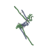
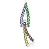
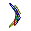
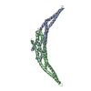
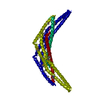
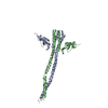
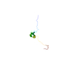
登録情報 データベース : PDB / ID : 3mudタイトル Structure of the Tropomyosin Overlap Complex from Chicken Smooth Muscle DNA repair protein XRCC4,Tropomyosin alpha-1 chain Tropomyosin alpha-1 chain,Microtubule-associated protein RP/EB family member 1 キーワード / / / 機能・相同性 分子機能 ドメイン・相同性 構成要素
/ / / / / / / / / / / / / / / / / / / / / / / / / / / / / / / / / / / / / / / / / / / / / / / / / / / / / / / / / / / / / / / / / / / / / / / / / / / / / / / / / / / / / / / / / / / / / / / / / / / / / / / / / / / / / / / / / / / / / / / / / / / / / / / / / / / / / / / / / / / / / / / / / / / / / / 生物種 Homo sapiens (ヒト)Gallus gallus (ニワトリ)手法 / / / 解像度 : 2.2 Å データ登録者 Klenchin, V.A. / Frye, J. / Rayment, I. ジャーナル : Biochemistry / 年 : 2010タイトル : Structure of the tropomyosin overlap complex from chicken smooth muscle: insight into the diversity of N-terminal recognition .著者 : Frye, J. / Klenchin, V.A. / Rayment, I. 履歴 登録 2010年5月2日 登録サイト / 処理サイト 改定 1.0 2010年6月23日 Provider / タイプ 改定 1.1 2011年7月13日 Group 改定 1.2 2017年5月31日 Group 改定 1.3 2023年9月6日 Group Data collection / Database references ... Data collection / Database references / Derived calculations / Refinement description カテゴリ chem_comp_atom / chem_comp_bond ... chem_comp_atom / chem_comp_bond / database_2 / pdbx_initial_refinement_model / struct_site Item _database_2.pdbx_DOI / _database_2.pdbx_database_accession ... _database_2.pdbx_DOI / _database_2.pdbx_database_accession / _struct_site.pdbx_auth_asym_id / _struct_site.pdbx_auth_comp_id / _struct_site.pdbx_auth_seq_id
すべて表示 表示を減らす
 ムービー
ムービー コントローラー


 データを開く
 基本情報
基本情報 要素
要素 キーワード
キーワード 機能・相同性情報
 Homo sapiens (ヒト)
 X線回折 /
  データ登録者
 引用
引用 構造の表示
構造の表示 ダウンロードとリンク
 その他のダウンロード
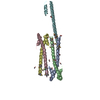

 PDBj
PDBj


 集合体
集合体


 分子量: 62.068 Da / 分子数: 1 / 由来タイプ: 合成 / 式: C2H6O2
 分子量: 96.063 Da / 分子数: 1 / 由来タイプ: 合成 / 式: SO4
 分子量: 18.015 Da / 分子数: 398 / 由来タイプ: 天然 / 式: H2O
 試料調製
試料調製 / ビームライン: 19-BM / 波長: 0.9793 Å
 解析
解析