Mass: 18.015 Da / Num. of mol.: 146 / Source method: isolated from a natural source / Formula: H2O
Compound details
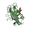
THE LIGAND DENOTED AS PGT HAS BEEN CO-PURIFIED ALONG WITH THE PROTEIN AND ITS EXACT IDENTITY IS NOT ...THE LIGAND DENOTED AS PGT HAS BEEN CO-PURIFIED ALONG WITH THE PROTEIN AND ITS EXACT IDENTITY IS NOT KNOWN. PGT REPRESENTS A CLOSE MATCH
Has protein modification
Y
-
Experimental details
-
Experiment
Experiment
Method: X-RAY DIFFRACTION / Number of used crystals: 1
-
Sample preparation
Crystal
Density Matthews: 2.29 Å3/Da / Density % sol: 46.29 %
Crystal grow
Temperature: 296 K / Method: vapor diffusion, sitting drop / pH: 7 Details: 18% PEG 4000, 100 mM Hepes pH 7.0, VAPOR DIFFUSION, SITTING DROP, temperature 296K
Resolution: 2→2.07 Å / Redundancy: 5.8 % / Rmerge(I) obs: 0.497 / Mean I/σ(I) obs: 4.66 / Num. unique all: 2077 / % possible all: 97.3
-
Processing
Software
Name
Version
Classification
HKL-2000
datacollection
SHELXS
phasing
REFMAC
5.5.0070
refinement
HKL-2000
datareduction
HKL-2000
datascaling
Refinement
Method to determine structure: SIRAS / Resolution: 2→30 Å / Cor.coef. Fo:Fc: 0.956 / Cor.coef. Fo:Fc free: 0.952 / SU B: 12.312 / SU ML: 0.149 / Isotropic thermal model: isotropic / Cross valid method: THROUGHOUT / ESU R Free: 0.176 / Stereochemistry target values: MAXIMUM LIKELIHOOD Details: A PEG4000 FRAGMENT (PGE) FROM CRYSTALLIZATION SOLUTION AND A GLYCEROL MOLECULE (GOL) FROM CRYO SOLUTION ARE MODELED.
Rfactor
Num. reflection
% reflection
Selection details
Rfree
0.25182
1095
5 %
RANDOM
Rwork
0.22119
-
-
-
obs
0.22275
20652
99.35 %
-
Solvent computation
Ion probe radii: 0.8 Å / Shrinkage radii: 0.8 Å / VDW probe radii: 1.4 Å / Solvent model: MASK
Displacement parameters
Biso mean: 37 Å2
Baniso -1
Baniso -2
Baniso -3
1-
0.34 Å2
0 Å2
0 Å2
2-
-
0.95 Å2
0 Å2
3-
-
-
-1.29 Å2
Refinement step
Cycle: LAST / Resolution: 2→30 Å
Protein
Nucleic acid
Ligand
Solvent
Total
Num. atoms
2153
0
110
146
2409
Refine LS restraints
Refine-ID
Type
Dev ideal
Dev ideal target
Number
X-RAY DIFFRACTION
r_bond_refined_d
0.017
0.022
2317
X-RAY DIFFRACTION
r_bond_other_d
0.005
0.02
1624
X-RAY DIFFRACTION
r_angle_refined_deg
1.723
2.008
3123
X-RAY DIFFRACTION
r_angle_other_deg
0.916
3.016
3786
X-RAY DIFFRACTION
r_dihedral_angle_1_deg
6.61
5
278
X-RAY DIFFRACTION
r_dihedral_angle_2_deg
31.11
24.444
90
X-RAY DIFFRACTION
r_dihedral_angle_3_deg
13.266
15
375
X-RAY DIFFRACTION
r_dihedral_angle_4_deg
10.48
15
9
X-RAY DIFFRACTION
r_chiral_restr
0.098
0.2
354
X-RAY DIFFRACTION
r_gen_planes_refined
0.006
0.021
2448
X-RAY DIFFRACTION
r_gen_planes_other
0.002
0.02
435
X-RAY DIFFRACTION
r_mcbond_it
0.886
1.5
1387
X-RAY DIFFRACTION
r_mcbond_other
0.225
1.5
562
X-RAY DIFFRACTION
r_mcangle_it
1.679
2
2244
X-RAY DIFFRACTION
r_scbond_it
2.42
3
930
X-RAY DIFFRACTION
r_scangle_it
3.965
4.5
878
Refine LS restraints NCS
Auth asym-ID: A / Refine-ID: X-RAY DIFFRACTION
Ens-ID
Dom-ID
Number
Type
Rms dev position (Å)
Weight position
1
1
802
tightpositional
0.04
0.05
2
2
109
mediumpositional
0.43
0.5
1
1
944
loosepositional
0.04
5
1
1
802
tightthermal
0.12
0.5
2
2
109
mediumthermal
0.25
2
1
1
944
loosethermal
0.13
10
LS refinement shell
Resolution: 2→2.052 Å / Total num. of bins used: 20
Rfactor
Num. reflection
% reflection
Rfree
0.341
65
-
Rwork
0.29
1444
-
obs
-
-
96.85 %
Refinement TLS params.
Method: refined / Refine-ID: X-RAY DIFFRACTION
ID
L11 (°2)
L12 (°2)
L13 (°2)
L22 (°2)
L23 (°2)
L33 (°2)
S11 (Å °)
S12 (Å °)
S13 (Å °)
S21 (Å °)
S22 (Å °)
S23 (Å °)
S31 (Å °)
S32 (Å °)
S33 (Å °)
T11 (Å2)
T12 (Å2)
T13 (Å2)
T22 (Å2)
T23 (Å2)
T33 (Å2)
Origin x (Å)
Origin y (Å)
Origin z (Å)
1
3.1501
-0.381
-0.3873
1.9341
1.197
2.573
0.0765
0.1117
-0.0612
0.0005
-0.1244
0.0079
-0.1019
-0.271
0.0479
0.0159
-0.0171
0.0044
0.5087
0.0155
0.2007
12.378
33.346
21.571
2
3.399
1.7343
-0.3644
3.7946
-0.819
11.9989
-0.0907
0.2865
-0.0746
0.1752
0.2543
-0.3486
-0.7914
0.2686
-0.1636
0.0718
-0.0271
-0.0061
0.4933
-0.023
0.3066
30.519
32.502
25.222
3
4.0954
-0.2251
1.4621
2.6053
0.6348
3.6825
0.0483
0.3628
0.0677
-0.171
-0.0544
-0.1268
-0.3759
-0.062
0.0061
0.0597
-0.0084
0.0371
0.4403
-0.011
0.192
17.63
36.875
22.2
4
10.1323
-2.386
2.0454
5.1584
-0.6746
3.9237
0.1747
0.4116
-0.0282
-0.1919
-0.143
-0.0047
-0.1359
-0.2527
-0.0317
0.0379
-0.0181
0.0536
0.4491
-0.0135
0.1602
13.586
30.194
18.343
5
3.5613
0.4822
-0.8243
1.9737
-1.2113
2.2123
0.1179
-0.2331
-0.0168
0.014
-0.205
0.0345
-0.1262
0.1598
0.087
0.032
0.0189
-0.0075
0.5421
-0.0539
0.178
27.69
32.956
-8.62
6
2.2578
-2.4176
-0.7269
4.5399
1.1069
11.2282
-0.0032
-0.4794
0.1281
-0.1907
0.2623
0.319
-0.8321
0.0652
-0.2591
0.1017
0.0226
-0.0196
0.6364
-0.0271
0.2827
9.466
32.215
-12.281
7
3.9364
1.3409
0.345
1.8489
-0.3959
3.3656
0.3105
-0.4187
0.1517
0.2349
-0.1507
0.1172
-0.4119
0.0796
-0.1598
0.1016
0.0311
0.0159
0.4802
-0.0223
0.2211
22.358
36.635
-9.303
8
9.8664
2.6521
2.0466
5.4168
1.7071
5.0445
0.3015
-0.4998
0.0323
0.1992
-0.3271
0.0843
0.0057
0.1421
0.0256
0.0464
0.0198
0.0311
0.5248
-0.0485
0.1358
26.317
29.844
-5.363
Refinement TLS group
ID
Refine-ID
Refine TLS-ID
Auth asym-ID
Auth seq-ID
1
X-RAY DIFFRACTION
1
A
21 - 93
2
X-RAY DIFFRACTION
2
A
94 - 110
3
X-RAY DIFFRACTION
3
A
111 - 132
4
X-RAY DIFFRACTION
4
A
133 - 158
5
X-RAY DIFFRACTION
5
B
19 - 93
6
X-RAY DIFFRACTION
6
B
94 - 110
7
X-RAY DIFFRACTION
7
B
111 - 132
8
X-RAY DIFFRACTION
8
B
133 - 158
+
About Yorodumi
-
News
-
Feb 9, 2022. New format data for meta-information of EMDB entries
New format data for meta-information of EMDB entries
Version 3 of the EMDB header file is now the official format.
The previous official version 1.9 will be removed from the archive.
In the structure databanks used in Yorodumi, some data are registered as the other names, "COVID-19 virus" and "2019-nCoV". Here are the details of the virus and the list of structure data.
Jan 31, 2019. EMDB accession codes are about to change! (news from PDBe EMDB page)
EMDB accession codes are about to change! (news from PDBe EMDB page)
The allocation of 4 digits for EMDB accession codes will soon come to an end. Whilst these codes will remain in use, new EMDB accession codes will include an additional digit and will expand incrementally as the available range of codes is exhausted. The current 4-digit format prefixed with “EMD-” (i.e. EMD-XXXX) will advance to a 5-digit format (i.e. EMD-XXXXX), and so on. It is currently estimated that the 4-digit codes will be depleted around Spring 2019, at which point the 5-digit format will come into force.
The EM Navigator/Yorodumi systems omit the EMD- prefix.
Related info.:Q: What is EMD? / ID/Accession-code notation in Yorodumi/EM Navigator
Yorodumi is a browser for structure data from EMDB, PDB, SASBDB, etc.
This page is also the successor to EM Navigator detail page, and also detail information page/front-end page for Omokage search.
The word "yorodu" (or yorozu) is an old Japanese word meaning "ten thousand". "mi" (miru) is to see.
Related info.:EMDB / PDB / SASBDB / Comparison of 3 databanks / Yorodumi Search / Aug 31, 2016. New EM Navigator & Yorodumi / Yorodumi Papers / Jmol/JSmol / Function and homology information / Changes in new EM Navigator and Yorodumi
 Movie
Movie Controller


 Open data
 Basic information
 Components
 Keywords
 Function and homology information
 X-RAY DIFFRACTION /
  Authors
 Citation
 Structure visualization
 Downloads & links
 Other downloads


 PDBj
PDBj
 Assembly
 Trichoplusia ni (cabbage looper) / Strain (production host): Hi-5 insect cells / References: UniProt: Q90890
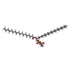
 Mass: 751.023 Da / Num. of mol.: 2 / Source method: obtained synthetically / Formula: C40H79O10P / Comment: phospholipid*YM
 Mass: 150.173 Da / Num. of mol.: 1 / Source method: obtained synthetically / Formula: C6H14O4
 Mass: 92.094 Da / Num. of mol.: 1 / Source method: obtained synthetically / Formula: C3H8O3
 Mass: 18.015 Da / Num. of mol.: 146 / Source method: isolated from a natural source / Formula: H2O
 Sample preparation
 / Beamline: BL11-1 / Wavelength: 0.9795 Å
 Processing