 Movie
Movie Controller

[English] 日本語
 Yorodumi
- PDB-3mrs: Crystal structure of shikimate kinase mutant (R57A) from Helicoba... -
+ Open data

- Basic information
| Entry | Database: PDB / ID: 3mrs | ||||||
|---|---|---|---|---|---|---|---|
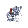
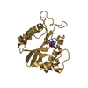
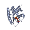
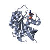
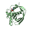
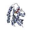
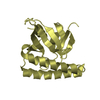
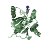
| Title | Crystal structure of shikimate kinase mutant (R57A) from Helicobacter pylori | ||||||
 Components Components | Shikimate kinase | ||||||
 Keywords Keywords | TRANSFERASE / helix-sheet complex | ||||||
| Function / homology |  Function and homology information Function and homology informationshikimate kinase / shikimate kinase activity / chorismate biosynthetic process / aromatic amino acid biosynthetic process / amino acid biosynthetic process / magnesium ion binding / ATP binding / cytosol Similarity search - Function | ||||||
| Biological species |   Helicobacter pylori (bacteria) Helicobacter pylori (bacteria) | ||||||
| Method |  X-RAY DIFFRACTION / SYNCHROTRON / MOLECULAR REPLACEMENT / Resolution: 2.4 Å X-RAY DIFFRACTION / SYNCHROTRON / MOLECULAR REPLACEMENT / Resolution: 2.4 Å | ||||||
 Authors Authors | Cheng, W.C. / Chen, T.J. / Lin, S.C. / Wang, W.C. | ||||||
 Citation Citation | Journal: Plos One / Year: 2012 Title: Structures of Helicobacter pylori shikimate kinase reveal a selective inhibitor-induced-fit mechanism Authors: Cheng, W.C. / Chen, Y.F. / Wang, H.J. / Hsu, K.C. / Lin, S.C. / Chen, T.J. / Yang, J.M. / Wang, W.C. | ||||||
| History |
|
- Structure visualization
| Structure viewer | Molecule: MolmilJmol/JSmol |
|---|
- Downloads & links
-Download
| PDBx/mmCIF format | 3mrs.cif.gz | 41.7 KB | Display | PDBx/mmCIF format |
|---|---|---|---|---|
| PDB format | pdb3mrs.ent.gz | 28.7 KB | Display | PDB format |
| PDBx/mmJSON format | 3mrs.json.gz | Tree view | PDBx/mmJSON format | |
| Others |  Other downloads Other downloads |
-Validation report
| Arichive directory | https://data.pdbj.org/pub/pdb/validation_reports/mr/3mrsftp://data.pdbj.org/pub/pdb/validation_reports/mr/3mrs | HTTPS FTP |
|---|
-Related structure data
| Related structure data |  3hr7C  3mufC  3n2eC  1zuiS S: Starting model for refinement C: citing same article ( |
|---|---|
| Similar structure data |


-Links
 PDBj
PDBj- Assembly
| Deposited unit | 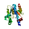
| ||||||||
|---|---|---|---|---|---|---|---|---|---|
| 1 |
| ||||||||
| Unit cell |
|
-Components
| #1: Protein | Mass: 19179.221 Da / Num. of mol.: 1 / Mutation: R57A Source method: isolated from a genetically manipulated source Source: (gene. exp.) Helicobacter pylori (bacteria) / Strain: 26695 / Gene: AROK / Plasmid: PQE30 / Production host: |
|---|---|
| #2: Water | ChemComp-HOH /  Mass: 18.015 Da / Num. of mol.: 25 / Source method: isolated from a natural source / Formula: H2O Mass: 18.015 Da / Num. of mol.: 25 / Source method: isolated from a natural source / Formula: H2O |
-Experimental details
-Experiment
| Experiment | Method: X-RAY DIFFRACTION / Number of used crystals: 1 |
|---|
- Sample preparation
| Crystal | Density Matthews: 2.15 Å3/Da / Density % sol: 42.7 % |
|---|---|
| Crystal grow | Temperature: 293 K / Method: vapor diffusion, hanging drop / pH: 8 Details: 18% PEG 4000, 8% isopropanol, 0.1M sodium HEPES, pH 8.0, VAPOR DIFFUSION, HANGING DROP, temperature 293K |
-Data collection
| Diffraction | Mean temperature: 193 K |
|---|---|
| Diffraction source | Source: SYNCHROTRON / Site: SPring-8  / Beamline: BL12B2 / Wavelength: 1 Å / Beamline: BL12B2 / Wavelength: 1 Å |
| Detector | Type: ADSC QUANTUM 210r / Detector: CCD / Date: Apr 10, 2010 |
| Radiation | Protocol: SINGLE WAVELENGTH / Monochromatic (M) / Laue (L): M / Scattering type: x-ray |
| Radiation wavelength | Wavelength: 1 Å / Relative weight: 1 |
| Reflection | Resolution: 2.4→30 Å / Num. obs: 6651 / % possible obs: 99.8 % / Observed criterion σ(F): 2 / Observed criterion σ(I): 2 / Redundancy: 10.4 % / Biso Wilson estimate: 44.2 Å2 / Rmerge(I) obs: 0.038 / Rsym value: 0.038 / Net I/σ(I): 56.02 |
| Reflection shell | Resolution: 2.4→2.49 Å / Redundancy: 10.6 % / Rmerge(I) obs: 0.131 / Mean I/σ(I) obs: 19.49 / Num. unique all: 1222 / Rsym value: 0.131 / % possible all: 100 |
- Processing
| Software |
| |||||||||||||||||||||||||||||||||||||||||||||||||||||||||||||||||
|---|---|---|---|---|---|---|---|---|---|---|---|---|---|---|---|---|---|---|---|---|---|---|---|---|---|---|---|---|---|---|---|---|---|---|---|---|---|---|---|---|---|---|---|---|---|---|---|---|---|---|---|---|---|---|---|---|---|---|---|---|---|---|---|---|---|---|
| Refinement | Method to determine structure: MOLECULAR REPLACEMENT Starting model: PDB ENTRY 1ZUI Resolution: 2.4→30 Å / Cor.coef. Fo:Fc: 0.899 / Cor.coef. Fo:Fc free: 0.869 / Isotropic thermal model: Isotropic / Cross valid method: THROUGHOUT / σ(F): 2 / σ(I): 2 / ESU R: 0.537 / ESU R Free: 0.291 / Stereochemistry target values: MAXIMUM LIKELIHOOD / Details: HYDROGENS HAVE BEEN ADDED IN THE RIDING POSITIONS
| |||||||||||||||||||||||||||||||||||||||||||||||||||||||||||||||||
| Solvent computation | Ion probe radii: 0.8 Å / Shrinkage radii: 0.8 Å / VDW probe radii: 1.4 Å / Solvent model: MASK | |||||||||||||||||||||||||||||||||||||||||||||||||||||||||||||||||
| Displacement parameters | Biso mean: 28.24 Å2
| |||||||||||||||||||||||||||||||||||||||||||||||||||||||||||||||||
| Refinement step | Cycle: LAST / Resolution: 2.4→30 Å
| |||||||||||||||||||||||||||||||||||||||||||||||||||||||||||||||||
| Refine LS restraints |
| |||||||||||||||||||||||||||||||||||||||||||||||||||||||||||||||||
| LS refinement shell | Resolution: 2.399→2.461 Å / Total num. of bins used: 20
|
