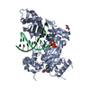
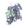
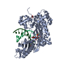
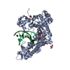
- PDB-3mr3: Human DNA polymerase eta - DNA ternary complex with the 3'T of a ... -
+
Open data
ID or keywords:
Loading...
-
Basic information
Entry
Database: PDB / ID: 3mr3
Title
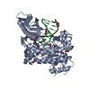
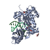
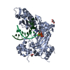
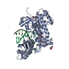
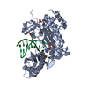
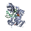
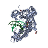
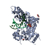
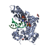
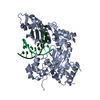
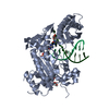
Human DNA polymerase eta - DNA ternary complex with the 3'T of a CPD in the active site (TT1)
Components
DNA (5'-D(*CP*AP*(TTD)P*AP*TP*GP*AP*CP*GP*CP*T)-3')
DNA (5'-D(*CP*AP*GP*CP*GP*TP*CP*AP*T)-3')
DNA polymerase eta
Keywords
TRANSFERASE/DNA / Pol eta / Polymerase / thymine dimer / CPD / XPV / Xeroderma pigmentosum variant / DNA damage / TRANSFERASE-DNA complex
Function / homology
Function and homology information
response to UV-C / error-free translesion synthesis / DNA synthesis involved in DNA repair / cellular response to UV-C / pyrimidine dimer repair / error-prone translesion synthesis / regulation of DNA repair / replication fork / Termination of translesion DNA synthesis / response to radiation ...response to UV-C / error-free translesion synthesis / DNA synthesis involved in DNA repair / cellular response to UV-C / pyrimidine dimer repair / error-prone translesion synthesis / regulation of DNA repair / replication fork / Termination of translesion DNA synthesis / response to radiation / Translesion Synthesis by POLH / HDR through Homologous Recombination (HRR) / site of double-strand break / DNA-directed DNA polymerase / damaged DNA binding / DNA-directed DNA polymerase activity / DNA replication / DNA repair / zinc ion binding / nucleoplasm / nucleus / cytosol Similarity search - Function
Ubiquitin-Binding Zinc Finger / DNApol eta/Rev1, HhH motif / DNA polymerase eta, ubiquitin-binding zinc finger / : / Zinc finger UBZ3-type profile. / DNA polymerase, Y-family, little finger domain / MutS, DNA mismatch repair protein, domain I - #60 / MutS, DNA mismatch repair protein, domain I / DNA polymerase, Y-family, little finger domain / impB/mucB/samB family C-terminal domain ...Ubiquitin-Binding Zinc Finger / DNApol eta/Rev1, HhH motif / DNA polymerase eta, ubiquitin-binding zinc finger / : / Zinc finger UBZ3-type profile. / DNA polymerase, Y-family, little finger domain / MutS, DNA mismatch repair protein, domain I - #60 / MutS, DNA mismatch repair protein, domain I / DNA polymerase, Y-family, little finger domain / impB/mucB/samB family C-terminal domain / UmuC domain / DNA polymerase, Y-family, little finger domain superfamily / impB/mucB/samB family / UmuC domain profile. / Reverse transcriptase/Diguanylate cyclase domain / Dna Ligase; domain 1 / 5' to 3' exonuclease, C-terminal subdomain / DNA polymerase; domain 1 / Reverse transcriptase/Diguanylate cyclase domain / Alpha-Beta Plaits / DNA/RNA polymerase superfamily / 2-Layer Sandwich / Orthogonal Bundle / 3-Layer(aba) Sandwich / Mainly Alpha / Alpha Beta Similarity search - Domain/homology
In the structure databanks used in Yorodumi, some data are registered as the other names, "COVID-19 virus" and "2019-nCoV". Here are the details of the virus and the list of structure data.
Jan 31, 2019. EMDB accession codes are about to change! (news from PDBe EMDB page)
EMDB accession codes are about to change! (news from PDBe EMDB page)
The allocation of 4 digits for EMDB accession codes will soon come to an end. Whilst these codes will remain in use, new EMDB accession codes will include an additional digit and will expand incrementally as the available range of codes is exhausted. The current 4-digit format prefixed with “EMD-” (i.e. EMD-XXXX) will advance to a 5-digit format (i.e. EMD-XXXXX), and so on. It is currently estimated that the 4-digit codes will be depleted around Spring 2019, at which point the 5-digit format will come into force.
The EM Navigator/Yorodumi systems omit the EMD- prefix.
Related info.:Q: What is EMD? / ID/Accession-code notation in Yorodumi/EM Navigator
Yorodumi is a browser for structure data from EMDB, PDB, SASBDB, etc.
This page is also the successor to EM Navigator detail page, and also detail information page/front-end page for Omokage search.
The word "yorodu" (or yorozu) is an old Japanese word meaning "ten thousand". "mi" (miru) is to see.
Related info.:EMDB / PDB / SASBDB / Comparison of 3 databanks / Yorodumi Search / Aug 31, 2016. New EM Navigator & Yorodumi / Yorodumi Papers / Jmol/JSmol / Function and homology information / Changes in new EM Navigator and Yorodumi
 Movie
Movie Controller

 Yorodumi
 Open data
 Basic information
 Components
 Keywords
 Function and homology information
 Homo sapiens (human)
 X-RAY DIFFRACTION /
  Authors
 Citation
 Structure visualization
 Downloads & links
 Other downloads
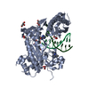


 PDBj
PDBj


 Assembly
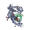


 Mass: 490.197 Da / Num. of mol.: 2 / Source method: obtained synthetically / Formula: C10H17N6O11P3
 Mass: 24.305 Da / Num. of mol.: 2 / Source method: obtained synthetically / Formula: Mg
 Mass: 92.094 Da / Num. of mol.: 6 / Source method: obtained synthetically / Formula: C3H8O3
 Sample preparation
 / Beamline: 22-ID / Wavelength: 1 Å
 Processing