 Movie
Movie Controller

[English] 日本語
 Yorodumi
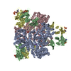
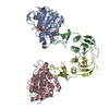
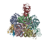
- PDB-3mpu: Crystal structure of the C47A/A241C disulfide-linked E. coli Aspa... -
+ Open data

- Basic information
| Entry | Database: PDB / ID: 3mpu | ||||||
|---|---|---|---|---|---|---|---|
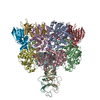
| Title | Crystal structure of the C47A/A241C disulfide-linked E. coli Aspartate Transcarbamoylase holoenzyme | ||||||
 Components Components |
| ||||||
 Keywords Keywords | TRANSFERASE / aspartate trancarbamoylase / disulfide bond / phosphate / catalysis / product release / ordered-sequential mechanism | ||||||
| Function / homology |  Function and homology information Function and homology informationaspartate carbamoyltransferase complex / pyrimidine nucleotide biosynthetic process / aspartate carbamoyltransferase / aspartate carbamoyltransferase activity / L-glutamine metabolic process / amino acid binding / protein homotrimerization / 'de novo' UMP biosynthetic process / 'de novo' pyrimidine nucleobase biosynthetic process / zinc ion binding ...aspartate carbamoyltransferase complex / pyrimidine nucleotide biosynthetic process / aspartate carbamoyltransferase / aspartate carbamoyltransferase activity / L-glutamine metabolic process / amino acid binding / protein homotrimerization / 'de novo' UMP biosynthetic process / 'de novo' pyrimidine nucleobase biosynthetic process / zinc ion binding / identical protein binding / cytosol / cytoplasm Similarity search - Function | ||||||
| Biological species |  | ||||||
| Method |  X-RAY DIFFRACTION / SYNCHROTRON / MOLECULAR REPLACEMENT / Resolution: 2.855 Å X-RAY DIFFRACTION / SYNCHROTRON / MOLECULAR REPLACEMENT / Resolution: 2.855 Å | ||||||
 Authors Authors | Mendes, K.R. / Kantrowitz, E.R. | ||||||
 Citation Citation | Journal: J.Mol.Biol. / Year: 2010 Title: The Pathway of Product Release from the R State of Aspartate Transcarbamoylase. Authors: Mendes, K.R. / Kantrowitz, E.R. | ||||||
| History |
|
- Structure visualization
| Structure viewer | Molecule: MolmilJmol/JSmol |
|---|
- Downloads & links
-Download
| PDBx/mmCIF format | 3mpu.cif.gz | 286.8 KB | Display | PDBx/mmCIF format |
|---|---|---|---|---|
| PDB format | pdb3mpu.ent.gz | 232.1 KB | Display | PDB format |
| PDBx/mmJSON format | 3mpu.json.gz | Tree view | PDBx/mmJSON format | |
| Others |  Other downloads Other downloads |
-Validation report
| Arichive directory | https://data.pdbj.org/pub/pdb/validation_reports/mp/3mpuftp://data.pdbj.org/pub/pdb/validation_reports/mp/3mpu | HTTPS FTP |
|---|
-Related structure data
| Related structure data | 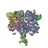 1d09S S: Starting model for refinement |
|---|---|
| Similar structure data |


-Links
 PDBj
PDBj
- Assembly
| Deposited unit | 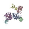
| |||||||||
|---|---|---|---|---|---|---|---|---|---|---|
| 1 | 
| |||||||||
| 2 | x 6
| |||||||||
| Unit cell |
| |||||||||
| Components on special symmetry positions |
|
-Components
| #1: Protein | Mass: 34337.105 Da / Num. of mol.: 3 / Mutation: C47A, A241C Source method: isolated from a genetically manipulated source Source: (gene. exp.) #2: Protein | Mass: 17143.625 Da / Num. of mol.: 3 Source method: isolated from a genetically manipulated source Source: (gene. exp.) #3: Chemical | ChemComp-PO4 /   Mass: 94.971 Da / Num. of mol.: 6 / Source method: obtained synthetically / Formula: PO4 Mass: 94.971 Da / Num. of mol.: 6 / Source method: obtained synthetically / Formula: PO4#4: Chemical |   Mass: 65.409 Da / Num. of mol.: 3 / Source method: obtained synthetically / Formula: Zn Mass: 65.409 Da / Num. of mol.: 3 / Source method: obtained synthetically / Formula: Zn#5: Water | ChemComp-HOH / |  Mass: 18.015 Da / Num. of mol.: 734 / Source method: isolated from a natural source / Formula: H2O Mass: 18.015 Da / Num. of mol.: 734 / Source method: isolated from a natural source / Formula: H2OHas protein modification | Y | |
|---|
-Experimental details
-Experiment
| Experiment | Method: X-RAY DIFFRACTION / Number of used crystals: 1 |
|---|
- Sample preparation
| Crystal | Density Matthews: 3.14 Å3/Da / Density % sol: 60.87 % |
|---|---|
| Crystal grow | Temperature: 293 K / Method: microdialysis / pH: 5.9 Details: Protein at 10 mg/ml was dialyzed against solution containing 100 mM KH2PO4, 3mM NaN3, pH 5.9, MICRODIALYSIS, temperature 293K |
-Data collection
| Diffraction | Mean temperature: 100 K |
|---|---|
| Diffraction source | Source: SYNCHROTRON / Site: NSLS  / Beamline: X29A / Wavelength: 1.0809 Å / Beamline: X29A / Wavelength: 1.0809 Å |
| Detector | Type: ADSC QUANTUM 315 / Detector: CCD / Date: Jan 21, 2009 / Details: fiber-optic taper |
| Radiation | Monochromator: sagitally focused Si(111) / Protocol: SINGLE WAVELENGTH / Monochromatic (M) / Laue (L): M / Scattering type: x-ray |
| Radiation wavelength | Wavelength: 1.0809 Å / Relative weight: 1 |
| Reflection | Resolution: 2.85→30 Å / Num. obs: 45620 / % possible obs: 99.9 % / Observed criterion σ(F): 0 / Observed criterion σ(I): 0 / Redundancy: 13.9 % / Biso Wilson estimate: 63.2 Å2 / Rmerge(I) obs: 0.094 / Rsym value: 0.094 / Net I/σ(I): 10 |
| Reflection shell | Resolution: 2.85→2.95 Å / Redundancy: 99.9 % / Rmerge(I) obs: 0.51 / Mean I/σ(I) obs: 4.8 / Num. unique all: 633869 / Rsym value: 0.51 / % possible all: 99.9 |
- Processing
| Software |
| |||||||||||||||||||||||||||||||||||||||||||||||||||||||||||||||||||||||||||||||||||||||||||||||||||||||||||||||||||||||
|---|---|---|---|---|---|---|---|---|---|---|---|---|---|---|---|---|---|---|---|---|---|---|---|---|---|---|---|---|---|---|---|---|---|---|---|---|---|---|---|---|---|---|---|---|---|---|---|---|---|---|---|---|---|---|---|---|---|---|---|---|---|---|---|---|---|---|---|---|---|---|---|---|---|---|---|---|---|---|---|---|---|---|---|---|---|---|---|---|---|---|---|---|---|---|---|---|---|---|---|---|---|---|---|---|---|---|---|---|---|---|---|---|---|---|---|---|---|---|---|---|
| Refinement | Method to determine structure: MOLECULAR REPLACEMENT Starting model: PDB entry 1D09, chain A Resolution: 2.855→29.923 Å / SU ML: 1.43 / σ(F): 1.33 / Phase error: 24.27 / Stereochemistry target values: ML
| |||||||||||||||||||||||||||||||||||||||||||||||||||||||||||||||||||||||||||||||||||||||||||||||||||||||||||||||||||||||
| Solvent computation | Shrinkage radii: 0.9 Å / VDW probe radii: 1.11 Å / Solvent model: FLAT BULK SOLVENT MODEL / Bsol: 34.943 Å2 / ksol: 0.251 e/Å3 | |||||||||||||||||||||||||||||||||||||||||||||||||||||||||||||||||||||||||||||||||||||||||||||||||||||||||||||||||||||||
| Displacement parameters |
| |||||||||||||||||||||||||||||||||||||||||||||||||||||||||||||||||||||||||||||||||||||||||||||||||||||||||||||||||||||||
| Refine analyze | Luzzati coordinate error obs: 0.289 Å | |||||||||||||||||||||||||||||||||||||||||||||||||||||||||||||||||||||||||||||||||||||||||||||||||||||||||||||||||||||||
| Refinement step | Cycle: LAST / Resolution: 2.855→29.923 Å
| |||||||||||||||||||||||||||||||||||||||||||||||||||||||||||||||||||||||||||||||||||||||||||||||||||||||||||||||||||||||
| Refine LS restraints |
| |||||||||||||||||||||||||||||||||||||||||||||||||||||||||||||||||||||||||||||||||||||||||||||||||||||||||||||||||||||||
| LS refinement shell | Refine-ID: X-RAY DIFFRACTION
|
