 Movie
Movie Controller

[English] 日本語
 Yorodumi
- PDB-3mgg: Crystal Structure of Methyl Transferase from Methanosarcina mazei -
+ Open data

- Basic information
| Entry | Database: PDB / ID: 3mgg | ||||||
|---|---|---|---|---|---|---|---|
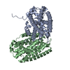
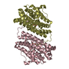
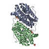
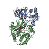
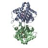
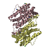
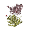
| Title | Crystal Structure of Methyl Transferase from Methanosarcina mazei | ||||||
 Components Components | Methyltransferase | ||||||
 Keywords Keywords | TRANSFERASE / NYSGXRC / PSI-II / Protein structure Initiative / structural genomics / New York SGX Research Center for Structural Genomics | ||||||
| Function / homology |  Function and homology information Function and homology informationTransferases; Transferring one-carbon groups; Methyltransferases / methyltransferase activity / methylation Similarity search - Function | ||||||
| Biological species |  Methanosarcina mazei (archaea) Methanosarcina mazei (archaea) | ||||||
| Method |  X-RAY DIFFRACTION / SYNCHROTRON / SAD / Resolution: 1.86 Å X-RAY DIFFRACTION / SYNCHROTRON / SAD / Resolution: 1.86 Å | ||||||
 Authors Authors | Syed Ibrahim, B. / Burley, S.K. / Swaminathan, S. / New York SGX Research Center for Structural Genomics (NYSGXRC) | ||||||
 Citation Citation | Journal: To be Published Title: Crystal Structure of Methyl Transferase from Methanosarcina mazei Authors: Syed Ibrahim, B. / Burley, S.K. / Swaminathan, S. | ||||||
| History |
|
- Structure visualization
| Structure viewer | Molecule: MolmilJmol/JSmol |
|---|
- Downloads & links
-Download
| PDBx/mmCIF format | 3mgg.cif.gz | 115.3 KB | Display | PDBx/mmCIF format |
|---|---|---|---|---|
| PDB format | pdb3mgg.ent.gz | 88.6 KB | Display | PDB format |
| PDBx/mmJSON format | 3mgg.json.gz | Tree view | PDBx/mmJSON format | |
| Others |  Other downloads Other downloads |
-Validation report
| Arichive directory | https://data.pdbj.org/pub/pdb/validation_reports/mg/3mggftp://data.pdbj.org/pub/pdb/validation_reports/mg/3mgg | HTTPS FTP |
|---|
-Related structure data
| Similar structure data | |
|---|---|
| Other databases |


-Links
 PDBj
PDBj- Assembly
| Deposited unit | 
| ||||||||
|---|---|---|---|---|---|---|---|---|---|
| 1 |
| ||||||||
| Unit cell |
|
-Components
| #1: Protein | Mass: 31463.078 Da / Num. of mol.: 2 Source method: isolated from a genetically manipulated source Source: (gene. exp.) Methanosarcina mazei (archaea) / Gene: MM_1949 / Plasmid: BC - pSGX3 (BC) / Production host:  References: UniProt: Q8PVL4, Transferases; Transferring one-carbon groups; Methyltransferases #2: Water | ChemComp-HOH / |  Mass: 18.015 Da / Num. of mol.: 312 / Source method: isolated from a natural source / Formula: H2O Mass: 18.015 Da / Num. of mol.: 312 / Source method: isolated from a natural source / Formula: H2OHas protein modification | Y | |
|---|
-Experimental details
-Experiment
| Experiment | Method: X-RAY DIFFRACTION / Number of used crystals: 1 |
|---|
- Sample preparation
| Crystal | Density Matthews: 2.46 Å3/Da / Density % sol: 50.04 % |
|---|---|
| Crystal grow | Temperature: 298 K / Method: vapor diffusion, sitting drop / pH: 4.5 Details: 0.1 M sodium acetate, 25% Peg 3350, pH 4.5, VAPOR DIFFUSION, SITTING DROP, temperature 298K |
-Data collection
| Diffraction | Mean temperature: 100 K |
|---|---|
| Diffraction source | Source: SYNCHROTRON / Site: NSLS  / Beamline: X29A / Wavelength: 0.9789 Å / Beamline: X29A / Wavelength: 0.9789 Å |
| Detector | Type: ADSC QUANTUM 315r / Detector: CCD / Date: Mar 25, 2010 / Details: Mirrors |
| Radiation | Monochromator: Si III / Protocol: SINGLE WAVELENGTH / Monochromatic (M) / Laue (L): M / Scattering type: x-ray |
| Radiation wavelength | Wavelength: 0.9789 Å / Relative weight: 1 |
| Reflection | Resolution: 1.86→50 Å / Num. all: 44542 / Num. obs: 44542 / % possible obs: 84.1 % / Redundancy: 12.9 % / Biso Wilson estimate: 12.8 Å2 / Rmerge(I) obs: 0.1 / Net I/σ(I): 8.9 |
| Reflection shell | Resolution: 1.86→1.93 Å / Redundancy: 12.1 % / Rmerge(I) obs: 0.653 / Num. unique all: 3943 / % possible all: 75.9 |
- Processing
| Software |
| ||||||||||||||||||||||||||||||||||||
|---|---|---|---|---|---|---|---|---|---|---|---|---|---|---|---|---|---|---|---|---|---|---|---|---|---|---|---|---|---|---|---|---|---|---|---|---|---|
| Refinement | Method to determine structure: SAD / Resolution: 1.86→45.54 Å / Rfactor Rfree error: 0.005 / Data cutoff high absF: 87742.23 / Data cutoff low absF: 0 / Isotropic thermal model: RESTRAINED / Cross valid method: THROUGHOUT / σ(F): 0 / Stereochemistry target values: Engh & Huber
| ||||||||||||||||||||||||||||||||||||
| Solvent computation | Solvent model: FLAT MODEL / Bsol: 28.0945 Å2 / ksol: 0.324767 e/Å3 | ||||||||||||||||||||||||||||||||||||
| Displacement parameters | Biso mean: 22.9 Å2
| ||||||||||||||||||||||||||||||||||||
| Refine analyze |
| ||||||||||||||||||||||||||||||||||||
| Refinement step | Cycle: LAST / Resolution: 1.86→45.54 Å
| ||||||||||||||||||||||||||||||||||||
| Refine LS restraints |
| ||||||||||||||||||||||||||||||||||||
| Refine LS restraints NCS | NCS model details: NONE | ||||||||||||||||||||||||||||||||||||
| LS refinement shell | Resolution: 1.86→1.98 Å / Rfactor Rfree error: 0.016 / Total num. of bins used: 6
| ||||||||||||||||||||||||||||||||||||
| Xplor file |
|
