Mass: 18.015 Da / Num. of mol.: 34 / Source method: isolated from a natural source / Formula: H2O
-
Experimental details
-
Experiment
Experiment
Method: X-RAY DIFFRACTION / Number of used crystals: 1
-
Sample preparation
Crystal
Density Matthews: 2.6 Å3/Da / Density % sol: 52.62 %
Crystal grow
Temperature: 295 K / Method: vapor diffusion / pH: 6.5 Details: Protein concentration of 7.5 mg/ml. 1:1 ul ratio of protein to crystallization solution. 1.0 M Sodium Citrate, 0.1 M Sodium Cacodylate, pH 6.5, Silver Bullet Reagent 29 (Hampton), VAPOR ...Details: Protein concentration of 7.5 mg/ml. 1:1 ul ratio of protein to crystallization solution. 1.0 M Sodium Citrate, 0.1 M Sodium Cacodylate, pH 6.5, Silver Bullet Reagent 29 (Hampton), VAPOR DIFFUSION, temperature 295K
Resolution: 2.91→72.57 Å / Cor.coef. Fo:Fc: 0.921 / Cor.coef. Fo:Fc free: 0.849 / SU B: 31.101 / SU ML: 0.283 / Cross valid method: THROUGHOUT / ESU R: 5.584 / ESU R Free: 0.411 / Stereochemistry target values: MAXIMUM LIKELIHOOD / Details: HYDROGENS HAVE BEEN ADDED IN THE RIDING POSITIONS
Rfactor
Num. reflection
% reflection
Selection details
Rfree
0.27193
764
5 %
RANDOM
Rwork
0.21672
-
-
-
obs
0.21957
14562
94.16 %
-
Solvent computation
Ion probe radii: 0.8 Å / Shrinkage radii: 0.8 Å / VDW probe radii: 1.4 Å / Solvent model: MASK
Displacement parameters
Biso mean: 45.313 Å2
Baniso -1
Baniso -2
Baniso -3
1-
1.19 Å2
0 Å2
-0 Å2
2-
-
-1.02 Å2
0 Å2
3-
-
-
-0.18 Å2
Refinement step
Cycle: LAST / Resolution: 2.91→72.57 Å
Protein
Nucleic acid
Ligand
Solvent
Total
Num. atoms
3550
0
44
34
3628
Refine LS restraints
Refine-ID
Type
Dev ideal
Dev ideal target
Number
X-RAY DIFFRACTION
r_bond_refined_d
0.014
0.021
3673
X-RAY DIFFRACTION
r_bond_other_d
0.001
0.02
2384
X-RAY DIFFRACTION
r_angle_refined_deg
1.495
1.963
4991
X-RAY DIFFRACTION
r_angle_other_deg
0.935
3
5795
X-RAY DIFFRACTION
r_dihedral_angle_1_deg
6.911
5
474
X-RAY DIFFRACTION
r_dihedral_angle_2_deg
35.54
23.5
140
X-RAY DIFFRACTION
r_dihedral_angle_3_deg
17.271
15
518
X-RAY DIFFRACTION
r_dihedral_angle_4_deg
20.566
15
18
X-RAY DIFFRACTION
r_chiral_restr
0.076
0.2
555
X-RAY DIFFRACTION
r_gen_planes_refined
0.007
0.021
4149
X-RAY DIFFRACTION
r_gen_planes_other
0.002
0.02
761
X-RAY DIFFRACTION
r_mcbond_it
0.572
1.5
2386
X-RAY DIFFRACTION
r_mcbond_other
0.09
1.5
977
X-RAY DIFFRACTION
r_mcangle_it
1.071
2
3762
X-RAY DIFFRACTION
r_scbond_it
1.583
3
1287
X-RAY DIFFRACTION
r_scangle_it
2.626
4.5
1229
LS refinement shell
Resolution: 2.915→2.99 Å / Total num. of bins used: 20
Rfactor
Num. reflection
% reflection
Rfree
0.415
36
-
Rwork
0.25
926
-
obs
-
-
81.87 %
Refinement TLS params.
Method: refined / Refine-ID: X-RAY DIFFRACTION
ID
L11 (°2)
L12 (°2)
L13 (°2)
L22 (°2)
L23 (°2)
L33 (°2)
S11 (Å °)
S12 (Å °)
S13 (Å °)
S21 (Å °)
S22 (Å °)
S23 (Å °)
S31 (Å °)
S32 (Å °)
S33 (Å °)
T11 (Å2)
T12 (Å2)
T13 (Å2)
T22 (Å2)
T23 (Å2)
T33 (Å2)
Origin x (Å)
Origin y (Å)
Origin z (Å)
1
4.5149
0.9351
-3.9253
2.7853
0.2797
11.7304
-0.0507
0.0861
0.311
-0.1488
0.031
-0.0984
-0.0941
-0.1968
0.0197
0.0155
0.0177
-0.0127
0.2696
0.0116
0.1468
-43.7636
30.0578
37.8445
2
3.0786
-0.5259
-1.3882
1.3974
0.4631
10.8683
0.12
-0.0409
-0.1122
-0.1555
0.0995
-0.1098
0.3157
-0.058
-0.2196
0.0816
0.0131
0.0155
0.1074
-0.0086
0.1055
-4.5657
19.6958
17.9434
3
3.5107
-0.1075
-0.2143
2.61
-0.3396
1.6082
-0.0405
-0.1267
-0.2779
-0.0602
0.0731
-0.0554
0.0539
-0.0945
-0.0326
0.0479
0.0076
-0.0068
0.167
0.0334
0.0732
-21.216
25.5375
33.2847
4
4.3712
1.5804
2.6149
6.8837
5.3336
8.142
0.0865
0.1473
-0.0616
-0.3229
0.2945
-0.1044
-0.349
-0.2532
-0.381
0.2186
0.0623
-0.0237
0.1254
0.0419
0.0558
-36.28
52.8257
5.5499
5
2.0868
-0.7441
0.4433
2.8297
-2.3276
3.2793
-0.1242
-0.193
0.3879
0.2904
-0.0968
-0.3454
-0.668
0.1414
0.221
0.3473
-0.0293
-0.0278
0.0676
-0.0852
0.1916
-18.8106
58.6401
20.6312
Refinement TLS group
ID
Refine-ID
Refine TLS-ID
Auth asym-ID
Auth seq-ID
1
X-RAY DIFFRACTION
1
A
-31 - -10
2
X-RAY DIFFRACTION
1
A
1 - 287
3
X-RAY DIFFRACTION
2
A
91 - 129
4
X-RAY DIFFRACTION
2
A
251 - 285
5
X-RAY DIFFRACTION
3
A
0 - 90
6
X-RAY DIFFRACTION
3
A
136 - 203
7
X-RAY DIFFRACTION
3
A
3 - 288
8
X-RAY DIFFRACTION
3
A
1 - 289
9
X-RAY DIFFRACTION
3
A
1 - 290
10
X-RAY DIFFRACTION
4
B
91 - 128
11
X-RAY DIFFRACTION
4
B
247 - 285
12
X-RAY DIFFRACTION
5
B
1 - 90
13
X-RAY DIFFRACTION
5
B
137 - 210
+
About Yorodumi
-
News
-
Feb 9, 2022. New format data for meta-information of EMDB entries
New format data for meta-information of EMDB entries
Version 3 of the EMDB header file is now the official format.
The previous official version 1.9 will be removed from the archive.
In the structure databanks used in Yorodumi, some data are registered as the other names, "COVID-19 virus" and "2019-nCoV". Here are the details of the virus and the list of structure data.
Jan 31, 2019. EMDB accession codes are about to change! (news from PDBe EMDB page)
EMDB accession codes are about to change! (news from PDBe EMDB page)
The allocation of 4 digits for EMDB accession codes will soon come to an end. Whilst these codes will remain in use, new EMDB accession codes will include an additional digit and will expand incrementally as the available range of codes is exhausted. The current 4-digit format prefixed with “EMD-” (i.e. EMD-XXXX) will advance to a 5-digit format (i.e. EMD-XXXXX), and so on. It is currently estimated that the 4-digit codes will be depleted around Spring 2019, at which point the 5-digit format will come into force.
The EM Navigator/Yorodumi systems omit the EMD- prefix.
Related info.:Q: What is EMD? / ID/Accession-code notation in Yorodumi/EM Navigator
Yorodumi is a browser for structure data from EMDB, PDB, SASBDB, etc.
This page is also the successor to EM Navigator detail page, and also detail information page/front-end page for Omokage search.
The word "yorodu" (or yorozu) is an old Japanese word meaning "ten thousand". "mi" (miru) is to see.
Related info.:EMDB / PDB / SASBDB / Comparison of 3 databanks / Yorodumi Search / Aug 31, 2016. New EM Navigator & Yorodumi / Yorodumi Papers / Jmol/JSmol / Function and homology information / Changes in new EM Navigator and Yorodumi
 Movie
Movie Controller


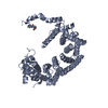
 Open data
 Basic information
 Components
 Keywords
 Function and homology information
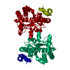
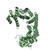
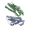
 uncultured soil bacterium (environmental samples)
 X-RAY DIFFRACTION /
  Authors
 Citation
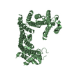
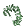
 Structure visualization
 Downloads & links
 Other downloads
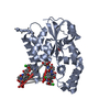


 PDBj
PDBj


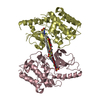
 Assembly

 Mass: 92.094 Da / Num. of mol.: 3 / Source method: obtained synthetically / Formula: C3H8O3
 Mass: 69.085 Da / Num. of mol.: 1 / Source method: obtained synthetically / Formula: C3H5N2
 Mass: 294.303 Da / Num. of mol.: 1 / Source method: obtained synthetically / Formula: C14H18N2O5
 Mass: 18.015 Da / Num. of mol.: 34 / Source method: isolated from a natural source / Formula: H2O
 Sample preparation
 / Beamline: X29A / Wavelength: 1.0809 Å
 Processing