Mass: 18.015 Da / Num. of mol.: 248 / Source method: isolated from a natural source / Formula: H2O
-
Experimental details
-
Experiment
Experiment
Method: X-RAY DIFFRACTION / Number of used crystals: 3
-
Sample preparation
Crystal
Density Matthews: 3.16 Å3/Da / Density % sol: 61.12 %
Crystal grow
Method: vapor diffusion / pH: 6.2 Details: 40% ethylenglycol 0.1 M Na/K/phosphate, pH 6.2, VAPOR DIFFUSION, temperature RTK
-
Data collection
Diffraction
ID
Mean temperature (K)
Crystal-ID
1
100
1
2
100
1
3
100
1
Diffraction source
Source
Site
Beamline
ID
Wavelength (Å)
SYNCHROTRON
SLS
X10SA
1
0.8266
SYNCHROTRON
SLS
X10SA
2
0.8266
SYNCHROTRON
APS
23-ID-B
3
1
Radiation
ID
Monochromator
Protocol
Monochromatic (M) / Laue (L)
Scattering type
Wavelength-ID
1
Doublecrystalmonochromator
SINGLEWAVELENGTH
M
x-ray
1
2
Doublecrystalmonochromator
SINGLEWAVELENGTH
M
x-ray
1
3
SINGLEWAVELENGTH
M
x-ray
1
Radiation wavelength
ID
Wavelength (Å)
Relative weight
1
0.8266
1
2
1
1
Reflection
Resolution: 1.27→50 Å / Num. all: 30089 / Num. obs: 25324 / % possible obs: 91.7 % / Observed criterion σ(I): 2 / Rmerge(I) obs: 0.06 / Net I/σ(I): 21.2
-
Processing
Software
Name
Classification
SHELXS
phasing
SHELXD
phasing
ACORN
phasing
SHELXL-97
refinement
HKL-2000
datareduction
HKL-2000
datascaling
Refinement
Method to determine structure: AB INITIO PHASING / Resolution: 1.27→18.51 Å / Cross valid method: thoughout / σ(F): 4 / σ(I): -3 / Details: conjugate gradient refinement against I
Rfactor
Num. reflection
Selection details
Rfree
0.22
1547
random
Rwork
0.179
-
-
obs
0.179
25324
-
all
-
29399
-
Refinement step
Cycle: LAST / Resolution: 1.27→18.51 Å
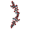
Protein
Nucleic acid
Ligand
Solvent
Total
Num. atoms
624
0
20
248
892
+
About Yorodumi
-
News
-
Feb 9, 2022. New format data for meta-information of EMDB entries
New format data for meta-information of EMDB entries
Version 3 of the EMDB header file is now the official format.
The previous official version 1.9 will be removed from the archive.
In the structure databanks used in Yorodumi, some data are registered as the other names, "COVID-19 virus" and "2019-nCoV". Here are the details of the virus and the list of structure data.
Jan 31, 2019. EMDB accession codes are about to change! (news from PDBe EMDB page)
EMDB accession codes are about to change! (news from PDBe EMDB page)
The allocation of 4 digits for EMDB accession codes will soon come to an end. Whilst these codes will remain in use, new EMDB accession codes will include an additional digit and will expand incrementally as the available range of codes is exhausted. The current 4-digit format prefixed with “EMD-” (i.e. EMD-XXXX) will advance to a 5-digit format (i.e. EMD-XXXXX), and so on. It is currently estimated that the 4-digit codes will be depleted around Spring 2019, at which point the 5-digit format will come into force.
The EM Navigator/Yorodumi systems omit the EMD- prefix.
Related info.:Q: What is EMD? / ID/Accession-code notation in Yorodumi/EM Navigator
Yorodumi is a browser for structure data from EMDB, PDB, SASBDB, etc.
This page is also the successor to EM Navigator detail page, and also detail information page/front-end page for Omokage search.
The word "yorodu" (or yorozu) is an old Japanese word meaning "ten thousand". "mi" (miru) is to see.
Related info.:EMDB / PDB / SASBDB / Comparison of 3 databanks / Yorodumi Search / Aug 31, 2016. New EM Navigator & Yorodumi / Yorodumi Papers / Jmol/JSmol / Function and homology information / Changes in new EM Navigator and Yorodumi
 Movie
Movie Controller


 Open data
 Basic information
 Components
 Keywords
 Function and homology information
 X-RAY DIFFRACTION /
  Authors
 Citation
 Structure visualization
 Downloads & links
 Other downloads
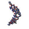
 PDBj
PDBj Assembly

 Mass: 62.068 Da / Num. of mol.: 5 / Source method: obtained synthetically / Formula: C2H6O2
 Mass: 18.015 Da / Num. of mol.: 248 / Source method: isolated from a natural source / Formula: H2O
 Sample preparation

 Processing