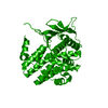
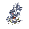
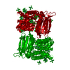
Mass: 41896.059 Da / Num. of mol.: 1 / Fragment: residues 188-512 Source method: isolated from a genetically manipulated source Details: The plasmid encodes a fusion protein comprising bacterial thioredoxin, a double His6-tag interspersed with a thrombin and an enterokinase cleavage site, and the CadC fragment comprising ...Details: The plasmid encodes a fusion protein comprising bacterial thioredoxin, a double His6-tag interspersed with a thrombin and an enterokinase cleavage site, and the CadC fragment comprising residues 188-512. Amino acid numbering of the crystal structure corresponds to the original numbering of the full length CadC protein sequence. Source: (gene. exp.) Escherichia coli (E. coli) / Strain: MG1655 / Gene: b4133, cadC, JW4094 / Plasmid: pET32a / Production host: Escherichia coli (E. coli) / Strain (production host): Origami B (DE3) pLysS / References: UniProt: P23890
#2: Chemical
ChemComp-RHE / rhenium (IV) hexachloride / hexachlororhenate ion
Mass: 398.925 Da / Num. of mol.: 16 / Source method: obtained synthetically / Formula: Cl6Re
Redundancy: 14.1 % / Av σ(I) over netI: 6.2 / Number: 247813 / Rmerge(I) obs: 0.095 / Rsym value: 0.095 / D res high: 2.3 Å / D res low: 69.768 Å / Num. obs: 17592 / % possible obs: 99
In the structure databanks used in Yorodumi, some data are registered as the other names, "COVID-19 virus" and "2019-nCoV". Here are the details of the virus and the list of structure data.
Jan 31, 2019. EMDB accession codes are about to change! (news from PDBe EMDB page)
EMDB accession codes are about to change! (news from PDBe EMDB page)
The allocation of 4 digits for EMDB accession codes will soon come to an end. Whilst these codes will remain in use, new EMDB accession codes will include an additional digit and will expand incrementally as the available range of codes is exhausted. The current 4-digit format prefixed with “EMD-” (i.e. EMD-XXXX) will advance to a 5-digit format (i.e. EMD-XXXXX), and so on. It is currently estimated that the 4-digit codes will be depleted around Spring 2019, at which point the 5-digit format will come into force.
The EM Navigator/Yorodumi systems omit the EMD- prefix.
Related info.:Q: What is EMD? / ID/Accession-code notation in Yorodumi/EM Navigator
Yorodumi is a browser for structure data from EMDB, PDB, SASBDB, etc.
This page is also the successor to EM Navigator detail page, and also detail information page/front-end page for Omokage search.
The word "yorodu" (or yorozu) is an old Japanese word meaning "ten thousand". "mi" (miru) is to see.
Related info.:EMDB / PDB / SASBDB / Comparison of 3 databanks / Yorodumi Search / Aug 31, 2016. New EM Navigator & Yorodumi / Yorodumi Papers / Jmol/JSmol / Function and homology information / Changes in new EM Navigator and Yorodumi
 Movie
Movie Controller

 Yorodumi
 Open data
 Basic information
 Components
 Keywords
 Function and homology information
 X-RAY DIFFRACTION /
  Authors
 Citation
 Structure visualization
 Downloads & links
 Other downloads
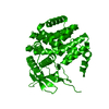


 PDBj
PDBj
 Assembly
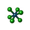
 Mass: 398.925 Da / Num. of mol.: 16 / Source method: obtained synthetically / Formula: Cl6Re
 Mass: 18.015 Da / Num. of mol.: 34 / Source method: isolated from a natural source / Formula: H2O
 Sample preparation
 / Beamline: 14.2 / Wavelength: 1.17652, 1.17705, 1.07813
 Processing