 Movie
Movie Controller

[English] 日本語
 Yorodumi
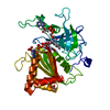
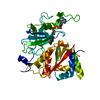
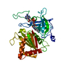
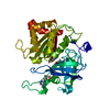
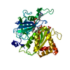
- PDB-3lvb: Crystal structure of the Ferredoxin:NADP+ reductase from maize ro... -
+ Open data

- Basic information
| Entry | Database: PDB / ID: 3lvb | ||||||
|---|---|---|---|---|---|---|---|
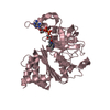
| Title | Crystal structure of the Ferredoxin:NADP+ reductase from maize root at 1.7 angstroms - Test Set Withheld | ||||||
 Components Components | Ferredoxin-NADP reductase | ||||||
 Keywords Keywords | OXIDOREDUCTASE / electron transport | ||||||
| Function / homology |  Function and homology information Function and homology informationoxidoreductase activity, acting on NAD(P)H, heme protein as acceptor / chloroplast thylakoid membrane protein complex / ferredoxin-NADP+ reductase / NADPH dehydrogenase activity / ferredoxin-NADP+ reductase activity / NADPH binding / photosynthesis / electron transport chain / electron transfer activity Similarity search - Function | ||||||
| Biological species |  | ||||||
| Method |  X-RAY DIFFRACTION / MOLECULAR REPLACEMENT / Resolution: 1.7 Å X-RAY DIFFRACTION / MOLECULAR REPLACEMENT / Resolution: 1.7 Å | ||||||
 Authors Authors | Faber, H.R. / Karplus, P.A. / Aliverti, A. / Ferioli, C. / Spinola, M. | ||||||
 Citation Citation | Journal: Biochemistry / Year: 2001 Title: Biochemical and crystallographic characterization of ferredoxin-NADP(+) reductase from nonphotosynthetic tissues Authors: Aliverti, A. / Faber, R. / Finnerty, C.M. / Ferioli, C. / Pandini, V. / Negri, A. / Karplus, P.A. / Zanetti, G. #1: Journal: To be PublishedTitle: Using a conformationally dependent stereochemical library improves refinement Authors: Tronrud, D.E. / Berkholz, D.S. / Karplus, P.A. | ||||||
| History |
|
- Structure visualization
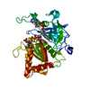
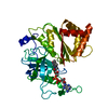
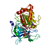
| Structure viewer | Molecule: MolmilJmol/JSmol |
|---|
- Downloads & links
-Download
| PDBx/mmCIF format | 3lvb.cif.gz | 82.2 KB | Display | PDBx/mmCIF format |
|---|---|---|---|---|
| PDB format | pdb3lvb.ent.gz | 59.7 KB | Display | PDB format |
| PDBx/mmJSON format | 3lvb.json.gz | Tree view | PDBx/mmJSON format | |
| Others |  Other downloads Other downloads |
-Validation report
| Arichive directory | https://data.pdbj.org/pub/pdb/validation_reports/lv/3lvbftp://data.pdbj.org/pub/pdb/validation_reports/lv/3lvb | HTTPS FTP |
|---|
-Related structure data
| Related structure data |  1jb9C  1fnbS C: citing same article ( S: Starting model for refinement |
|---|---|
| Similar structure data |


-Links
 PDBj
PDBj

- Assembly
| Deposited unit | 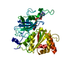
| ||||||||
|---|---|---|---|---|---|---|---|---|---|
| 1 |
| ||||||||
| Unit cell |
| ||||||||
| Details | The biological assembly is the contents of the asymmetric unit |
-Components
| #1: Protein | Mass: 34848.461 Da / Num. of mol.: 1 Source method: isolated from a genetically manipulated source Source: (gene. exp.)  |
|---|---|
| #2: Chemical | ChemComp-FAD /   Mass: 785.550 Da / Num. of mol.: 1 / Source method: obtained synthetically / Formula: C27H33N9O15P2 / Comment: FAD*YM Mass: 785.550 Da / Num. of mol.: 1 / Source method: obtained synthetically / Formula: C27H33N9O15P2 / Comment: FAD*YM |
| #3: Water | ChemComp-HOH /  Mass: 18.015 Da / Num. of mol.: 274 / Source method: isolated from a natural source / Formula: H2O Mass: 18.015 Da / Num. of mol.: 274 / Source method: isolated from a natural source / Formula: H2O |
| Has protein modification | Y |
-Experimental details
-Experiment
| Experiment | Method: X-RAY DIFFRACTION / Number of used crystals: 1 |
|---|
- Sample preparation
| Crystal | Density Matthews: 2.79 Å3/Da / Density % sol: 55.89 % |
|---|---|
| Crystal grow | Temperature: 298 K / Method: vapor diffusion, hanging drop / pH: 6 Details: PEG 8000, Na cacodylate, Mg Acetate, pH 6.0, VAPOR DIFFUSION, HANGING DROP, temperature 298K |
-Data collection
| Diffraction | Mean temperature: 298 K |
|---|---|
| Diffraction source | Source: ROTATING ANODE / Type: RIGAKU RU300 / Wavelength: 1.54 Å |
| Detector | Type: RIGAKU RAXIS IV / Detector: IMAGE PLATE / Date: Mar 1, 2000 / Details: Yale Mirrors |
| Radiation | Protocol: SINGLE WAVELENGTH / Monochromatic (M) / Laue (L): M / Scattering type: x-ray |
| Radiation wavelength | Wavelength: 1.54 Å / Relative weight: 1 |
| Reflection | Resolution: 1.65→50 Å / Num. all: 47556 / Num. obs: 47556 / % possible obs: 98.9 % / Observed criterion σ(F): 0 / Observed criterion σ(I): 0 / Redundancy: 5.4 % / Rmerge(I) obs: 0.064 / Net I/σ(I): 13.5 |
| Reflection shell | Resolution: 1.65→1.7 Å / Redundancy: 3.3 % / Rmerge(I) obs: 0.294 / Mean I/σ(I) obs: 3 / % possible all: 96.1 |
- Processing
| Software |
| |||||||||||||||||||||||||
|---|---|---|---|---|---|---|---|---|---|---|---|---|---|---|---|---|---|---|---|---|---|---|---|---|---|---|
| Refinement | Method to determine structure: MOLECULAR REPLACEMENT Starting model: 1FNB Resolution: 1.7→23.94 Å / Isotropic thermal model: isotropic / Cross valid method: throughhout / σ(F): 0 / σ(I): 0 / Stereochemistry target values: Engh & Huber
| |||||||||||||||||||||||||
| Displacement parameters |
| |||||||||||||||||||||||||
| Refinement step | Cycle: LAST / Resolution: 1.7→23.94 Å
| |||||||||||||||||||||||||
| Refine LS restraints |
|
