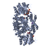
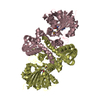
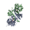
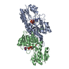
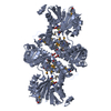
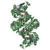
The biological unit is a dimer. There are half of 2 biological units in the asymmetric unit. The dimer generated from the monomer in the asymmetric unit by the operations: x, -y,-z and -x, y,-z+1/2
-
Components
#1: Protein
CalO1Methyltransferase
Mass: 37936.082 Da / Num. of mol.: 2 Source method: isolated from a genetically manipulated source Source: (gene. exp.) Micromonospora echinospora (bacteria) / Gene: calO1 / Plasmid: pET16b / Production host: Escherichia coli (E. coli) / Strain (production host): B834 P(RARE2) / References: UniProt: Q8KNE5
Mass: 18.015 Da / Num. of mol.: 181 / Source method: isolated from a natural source / Formula: H2O
Has protein modification
Y
-
Experimental details
-
Experiment
Experiment
Method: X-RAY DIFFRACTION / Number of used crystals: 1
-
Sample preparation
Crystal
Density Matthews: 2.36 Å3/Da / Density % sol: 47.89 %
Crystal grow
Temperature: 298 K / Method: vapor diffusion, hanging drop / pH: 7 Details: Protein Solution 20mg/ml CalO1 protein, 20mM Tris pH 8, mixed in a 1:1 ratio with the well solution 20% MEPEG 5K, 0.2M Glycine, 0.1M BTP pH 7.0. Cryoprotected with 20% ethylene glycol, 20% ...Details: Protein Solution 20mg/ml CalO1 protein, 20mM Tris pH 8, mixed in a 1:1 ratio with the well solution 20% MEPEG 5K, 0.2M Glycine, 0.1M BTP pH 7.0. Cryoprotected with 20% ethylene glycol, 20% MEPEG 5K, 0.2M Glycine, 0.1M BTP pH 7.0, VAPOR DIFFUSION, HANGING DROP, temperature 298K
Redundancy: 12.3 % / Av σ(I) over netI: 22.53 / Number: 344828 / Rmerge(I) obs: 0.12 / Χ2: 1.39 / D res high: 2.4 Å / D res low: 50 Å / Num. obs: 28057 / % possible obs: 97.7
Diffraction reflection shell
Highest resolution (Å)
Lowest resolution (Å)
% possible obs (%)
ID
Rmerge(I) obs
Chi squared
Redundancy
6.51
50
99.8
1
0.072
3.337
13.4
5.17
6.51
100
1
0.088
2.035
13.8
4.52
5.17
100
1
0.084
1.571
13.5
4.1
4.52
100
1
0.088
1.314
13.1
3.81
4.1
99.9
1
0.085
1.279
13
3.58
3.81
100
1
0.093
1.236
13.1
3.41
3.58
100
1
0.111
1.192
13
3.26
3.41
100
1
0.13
1.104
12.9
3.13
3.26
100
1
0.156
1.172
13.1
3.02
3.13
100
1
0.191
1.185
12.8
2.93
3.02
100
1
0.208
1.26
13
2.85
2.93
100
1
0.243
1.151
12.9
2.77
2.85
100
1
0.292
1.204
12.6
2.7
2.77
100
1
0.333
1.183
12.4
2.64
2.7
99.2
1
0.369
1.092
11.5
2.59
2.64
97.6
1
0.404
1.128
11.1
2.53
2.59
95.2
1
0.404
1.155
10.3
2.49
2.53
91.2
1
0.437
1.134
10.1
2.44
2.49
87.7
1
0.513
1.107
9.5
2.4
2.44
83.4
1
0.525
1.051
9
Reflection
Resolution: 2.4→50 Å / Num. obs: 28057 / % possible obs: 97.7 % / Redundancy: 12.3 % / Rmerge(I) obs: 0.12 / Χ2: 1.387 / Net I/σ(I): 9.7
 Movie
Movie Controller

 Yorodumi
 Open data
 Basic information
 Components
 Keywords
 Function and homology information
 Micromonospora echinospora (bacteria)
 X-RAY DIFFRACTION /
  Authors
 Citation
 Structure visualization
 Downloads & links
 Other downloads
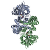

 PDBj
PDBj Assembly
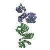

 Mass: 384.411 Da / Num. of mol.: 2 / Source method: obtained synthetically / Formula: C14H20N6O5S
 Mass: 62.068 Da / Num. of mol.: 2 / Source method: obtained synthetically / Formula: C2H6O2
 Mass: 18.015 Da / Num. of mol.: 181 / Source method: isolated from a natural source / Formula: H2O
 Sample preparation
 / Beamline: 23-ID-D / Wavelength: 0.9794, 0.9641