 Movie
Movie Controller

[English] 日本語
 Yorodumi
- PDB-3ls2: Crystal structure of an S-formylglutathione hydrolase from Pseudo... -
+ Open data

- Basic information
| Entry | Database: PDB / ID: 3ls2 | ||||||
|---|---|---|---|---|---|---|---|
| Title | Crystal structure of an S-formylglutathione hydrolase from Pseudoalteromonas haloplanktis TAC125 | ||||||
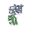
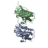
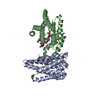
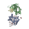
 Components Components | S-formylglutathione Hydrolase | ||||||
 Keywords Keywords | HYDROLASE / S-formylglutathione hydrolase / psychrophilic organism / Pseudoalteromonas haloplanktis | ||||||
| Function / homology |  Function and homology information Function and homology informationS-formylglutathione hydrolase / S-formylglutathione hydrolase activity / formaldehyde catabolic process / carboxylic ester hydrolase activity / cytosol Similarity search - Function | ||||||
| Biological species |  Pseudoalteromonas haloplanktis (bacteria) Pseudoalteromonas haloplanktis (bacteria) | ||||||
| Method |  X-RAY DIFFRACTION / MOLECULAR REPLACEMENT / Resolution: 2.2 Å X-RAY DIFFRACTION / MOLECULAR REPLACEMENT / Resolution: 2.2 Å | ||||||
 Authors Authors | Alterio, V. / De Simone, G. | ||||||
 Citation Citation | Journal: Biopolymers / Year: 2010 Title: Crystal structure of an S-formylglutathione hydrolase from Pseudoalteromonas haloplanktis TAC125. Authors: Alterio, V. / Aurilia, V. / Romanelli, A. / Parracino, A. / Saviano, M. / D'Auria, S. / De Simone, G. | ||||||
| History |
|
- Structure visualization
| Structure viewer | Molecule: MolmilJmol/JSmol |
|---|
- Downloads & links
-Download
| PDBx/mmCIF format | 3ls2.cif.gz | 243.6 KB | Display | PDBx/mmCIF format |
|---|---|---|---|---|
| PDB format | pdb3ls2.ent.gz | 194.9 KB | Display | PDB format |
| PDBx/mmJSON format | 3ls2.json.gz | Tree view | PDBx/mmJSON format | |
| Others |  Other downloads Other downloads |
-Validation report
| Summary document | 3ls2_validation.pdf.gz | 446 KB | Display | wwPDB validaton report |
|---|---|---|---|---|
| Full document | 3ls2_full_validation.pdf.gz | 455.2 KB | Display | |
| Data in XML | 3ls2_validation.xml.gz | 51.1 KB | Display | |
| Data in CIF | 3ls2_validation.cif.gz | 75 KB | Display | |
| Arichive directory | https://data.pdbj.org/pub/pdb/validation_reports/ls/3ls2ftp://data.pdbj.org/pub/pdb/validation_reports/ls/3ls2 | HTTPS FTP |
-Related structure data
| Related structure data | 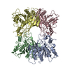 1pv1S S: Starting model for refinement |
|---|---|
| Similar structure data |


-Links
 PDBj
PDBj- Assembly
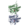
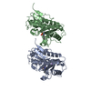
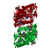
| Deposited unit | 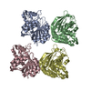
| ||||||||
|---|---|---|---|---|---|---|---|---|---|
| 1 | 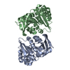
| ||||||||
| 2 | 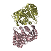
| ||||||||
| Unit cell |
|
-Components
| #1: Protein | Mass: 30915.732 Da / Num. of mol.: 4 Source method: isolated from a genetically manipulated source Source: (gene. exp.) Pseudoalteromonas haloplanktis (bacteria)Strain: TAC125 / Gene: PSHAa1385 / Plasmid: pET28a / Production host: References: UniProt: Q3IL66, Hydrolases; Acting on ester bonds; Carboxylic-ester hydrolases #2: Chemical | ChemComp-CL /   Mass: 35.453 Da / Num. of mol.: 4 / Source method: obtained synthetically / Formula: Cl Mass: 35.453 Da / Num. of mol.: 4 / Source method: obtained synthetically / Formula: Cl#3: Water | ChemComp-HOH / |  Mass: 18.015 Da / Num. of mol.: 1054 / Source method: isolated from a natural source / Formula: H2O Mass: 18.015 Da / Num. of mol.: 1054 / Source method: isolated from a natural source / Formula: H2O |
|---|
-Experimental details
-Experiment
| Experiment | Method: X-RAY DIFFRACTION / Number of used crystals: 1 |
|---|
- Sample preparation
| Crystal | Density Matthews: 1.98 Å3/Da / Density % sol: 37.94 % |
|---|---|
| Crystal grow | Temperature: 293 K / Method: vapor diffusion, hanging drop / pH: 7 Details: 20% (w/v) PEG MME 5000, 0.2 M sodium acetate, 0.1 M Tris-HCl, pH 7.0, VAPOR DIFFUSION, HANGING DROP, temperature 293 K |
-Data collection
| Diffraction | Mean temperature: 100 K |
|---|---|
| Diffraction source | Source: ROTATING ANODE / Type: RIGAKU MICROMAX-007 HF / Wavelength: 1.54178 Å |
| Detector | Type: RIGAKU SATURN 944 / Detector: CCD / Date: Jul 10, 2009 / Details: mirrors |
| Radiation | Protocol: SINGLE WAVELENGTH / Monochromatic (M) / Laue (L): M / Scattering type: x-ray |
| Radiation wavelength | Wavelength: 1.54178 Å / Relative weight: 1 |
| Reflection | Resolution: 2.2→20 Å / Num. all: 49990 / Num. obs: 49990 / % possible obs: 98.7 % / Redundancy: 3.9 % / Rmerge(I) obs: 0.053 / Net I/σ(I): 19.5 |
| Reflection shell | Resolution: 2.2→2.28 Å / Redundancy: 2.6 % / Rmerge(I) obs: 0.166 / Mean I/σ(I) obs: 5.9 / Num. unique all: 4666 / % possible all: 93.6 |
- Processing
| Software |
| ||||||||||||||||||||
|---|---|---|---|---|---|---|---|---|---|---|---|---|---|---|---|---|---|---|---|---|---|
| Refinement | Method to determine structure: MOLECULAR REPLACEMENT Starting model: PDB entry 1PV1 Resolution: 2.2→20 Å / Isotropic thermal model: isotropic / σ(F): 0 / Stereochemistry target values: Engh & Huber
| ||||||||||||||||||||
| Displacement parameters | Biso mean: 15.6 Å2 | ||||||||||||||||||||
| Refinement step | Cycle: LAST / Resolution: 2.2→20 Å
| ||||||||||||||||||||
| Refine LS restraints |
|
