 Movie
Movie Controller

[English] 日本語
 Yorodumi
- PDB-3lqu: Crystal structure of 3,4-Dihydroxy-2-butanone 4-phosphate synthas... -
+ Open data

- Basic information
| Entry | Database: PDB / ID: 3lqu | ||||||
|---|---|---|---|---|---|---|---|
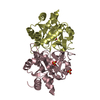
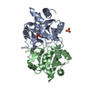
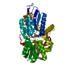
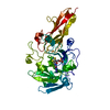
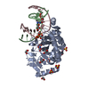
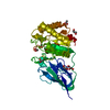
| Title | Crystal structure of 3,4-Dihydroxy-2-butanone 4-phosphate synthase complexed with Ribulose-5 phosphate | ||||||
 Components Components | 3,4-dihydroxy-2-butanone 4-phosphate synthase | ||||||
 Keywords Keywords | LYASE / DHBPS / R5P / ribB / Metal binding / Riboflavin biosynthesis | ||||||
| Function / homology |  Function and homology information Function and homology information3,4-dihydroxy-2-butanone-4-phosphate synthase / 3,4-dihydroxy-2-butanone-4-phosphate synthase activity / riboflavin biosynthetic process / manganese ion binding / magnesium ion binding / cytosol Similarity search - Function | ||||||
| Biological species |  Salmonella typhimurium (bacteria) Salmonella typhimurium (bacteria) | ||||||
| Method |  X-RAY DIFFRACTION / MOLECULAR REPLACEMENT / Resolution: 2.522 Å X-RAY DIFFRACTION / MOLECULAR REPLACEMENT / Resolution: 2.522 Å | ||||||
 Authors Authors | Kumar, P. / Karthikeyan, S. | ||||||
 Citation Citation | Journal: Proteins / Year: 2010 Title: Potential anti-bacterial drug target: structural characterization of 3,4-dihydroxy-2-butanone-4-phosphate synthase from Salmonella typhimurium LT2. Authors: Kumar, P. / Singh, M. / Gautam, R. / Karthikeyan, S. | ||||||
| History |
|
- Structure visualization
| Structure viewer | Molecule: MolmilJmol/JSmol |
|---|
- Downloads & links
-Download
| PDBx/mmCIF format | 3lqu.cif.gz | 88.8 KB | Display | PDBx/mmCIF format |
|---|---|---|---|---|
| PDB format | pdb3lqu.ent.gz | 67.3 KB | Display | PDB format |
| PDBx/mmJSON format | 3lqu.json.gz | Tree view | PDBx/mmJSON format | |
| Others |  Other downloads Other downloads |
-Validation report
| Arichive directory | https://data.pdbj.org/pub/pdb/validation_reports/lq/3lquftp://data.pdbj.org/pub/pdb/validation_reports/lq/3lqu | HTTPS FTP |
|---|
-Related structure data
| Related structure data |  3lrjC 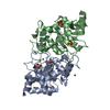 3ls6C 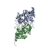 1g57S S: Starting model for refinement C: citing same article ( |
|---|---|
| Similar structure data |


-Links
 PDBj
PDBj- Assembly
| Deposited unit | 
| |||||||||
|---|---|---|---|---|---|---|---|---|---|---|
| 1 |
| |||||||||
| Unit cell |
| |||||||||
| Components on special symmetry positions |
|
-Components
| #1: Protein | Mass: 23335.439 Da / Num. of mol.: 2 Source method: isolated from a genetically manipulated source Source: (gene. exp.) Salmonella typhimurium (bacteria) / Strain: LT2 / Gene: ribB, STM3195 / Plasmid: pET28c / Production host: References: UniProt: P66032, 3,4-dihydroxy-2-butanone-4-phosphate synthase #2: Sugar | ChemComp-5SP / |   Type: saccharide / Mass: 230.110 Da / Num. of mol.: 1 Type: saccharide / Mass: 230.110 Da / Num. of mol.: 1Source method: isolated from a genetically manipulated source Formula: C5H11O8P #3: Water | ChemComp-HOH / |  Mass: 18.015 Da / Num. of mol.: 128 / Source method: isolated from a natural source / Formula: H2O Mass: 18.015 Da / Num. of mol.: 128 / Source method: isolated from a natural source / Formula: H2O |
|---|
-Experimental details
-Experiment
| Experiment | Method: X-RAY DIFFRACTION / Number of used crystals: 1 |
|---|
- Sample preparation
| Crystal | Density Matthews: 2.05 Å3/Da / Density % sol: 40.02 % |
|---|---|
| Crystal grow | Temperature: 293 K / Method: vapor diffusion, hanging drop / pH: 6.5 Details: 30% PEG 8000, 100mM MES-Na, 0.2M Ammonium Salt, pH 6.5, VAPOR DIFFUSION, HANGING DROP, temperature 293K |
-Data collection
| Diffraction | Mean temperature: 100 K |
|---|---|
| Diffraction source | Source: ROTATING ANODE / Type: RIGAKU MICROMAX-007 HF / Wavelength: 1.542 Å |
| Detector | Type: MAR scanner 345 mm plate / Detector: IMAGE PLATE / Date: Jul 18, 2009 / Details: Osmic VariMax |
| Radiation | Monochromator: Mirror / Protocol: SINGLE WAVELENGTH / Monochromatic (M) / Laue (L): M / Scattering type: x-ray |
| Radiation wavelength | Wavelength: 1.542 Å / Relative weight: 1 |
| Reflection | Resolution: 2.52→50 Å / Num. all: 12300 / Num. obs: 12279 / % possible obs: 92.3 % / Observed criterion σ(I): 2 / Redundancy: 2.5 % / Biso Wilson estimate: 29.6 Å2 / Rmerge(I) obs: 0.147 / Rsym value: 0.147 / Net I/σ(I): 7.85 |
| Reflection shell | Resolution: 2.52→2.61 Å / Redundancy: 2.1 % / Rmerge(I) obs: 0.487 / Mean I/σ(I) obs: 2.01 / Num. unique all: 1112 / Rsym value: 0.487 / % possible all: 85.6 |
- Processing
| Software |
| ||||||||||||||||||||||||||||||
|---|---|---|---|---|---|---|---|---|---|---|---|---|---|---|---|---|---|---|---|---|---|---|---|---|---|---|---|---|---|---|---|
| Refinement | Method to determine structure: MOLECULAR REPLACEMENT Starting model: 1G57 Resolution: 2.522→19.53 Å / SU ML: 0.41 / Isotropic thermal model: Isotropic / Cross valid method: THROUGHOUT / σ(F): 0.09 / Phase error: 27.57 / Stereochemistry target values: ML
| ||||||||||||||||||||||||||||||
| Solvent computation | Shrinkage radii: 0.9 Å / VDW probe radii: 1.11 Å / Solvent model: FLAT BULK SOLVENT MODEL / Bsol: 49.553 Å2 / ksol: 0.372 e/Å3 | ||||||||||||||||||||||||||||||
| Displacement parameters |
| ||||||||||||||||||||||||||||||
| Refine analyze | Luzzati coordinate error obs: 0.41 Å | ||||||||||||||||||||||||||||||
| Refinement step | Cycle: LAST / Resolution: 2.522→19.53 Å
| ||||||||||||||||||||||||||||||
| Refine LS restraints |
| ||||||||||||||||||||||||||||||
| LS refinement shell | Refine-ID: X-RAY DIFFRACTION / Total num. of bins used: 4
|
