 Movie
Movie Controller

[English] 日本語
 Yorodumi
- PDB-3lnb: Crystal Structure Analysis of Arylamine N-acetyltransferase C fro... -
+ Open data

- Basic information
| Entry | Database: PDB / ID: 3lnb | ||||||
|---|---|---|---|---|---|---|---|
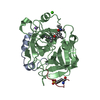
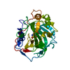
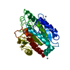
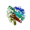
| Title | Crystal Structure Analysis of Arylamine N-acetyltransferase C from Bacillus anthracis | ||||||
 Components Components | N-acetyltransferase family protein | ||||||
 Keywords Keywords | TRANSFERASE / arylamine N-acetyltransferase / nat / acetyltransferase / Acyltransferase | ||||||
| Function / homology |  Function and homology information Function and homology information | ||||||
| Biological species |  | ||||||
| Method |  X-RAY DIFFRACTION / SYNCHROTRON / MOLECULAR REPLACEMENT / molecular replacement / Resolution: 2.01 Å X-RAY DIFFRACTION / SYNCHROTRON / MOLECULAR REPLACEMENT / molecular replacement / Resolution: 2.01 Å | ||||||
 Authors Authors | Li de la Sierra-Gallay, I. / Pluvinage, B. / Rodrigues-Lima, F. | ||||||
 Citation Citation | Journal: Febs Lett. / Year: 2011 Title: The Bacillus anthracis arylamine N-acetyltransferase ((BACAN)NAT1) that inactivates sulfamethoxazole, reveals unusual structural features compared with the other NAT isoenzymes. Authors: Pluvinage, B. / Li de la Sierra-Gallay, I. / Kubiak, X. / Xu, X. / Dairou, J. / Dupret, J.M. / Rodrigues-Lima, F. | ||||||
| History |
|
- Structure visualization
| Structure viewer | Molecule: MolmilJmol/JSmol |
|---|
- Downloads & links
-Download
| PDBx/mmCIF format | 3lnb.cif.gz | 71.6 KB | Display | PDBx/mmCIF format |
|---|---|---|---|---|
| PDB format | pdb3lnb.ent.gz | 50.3 KB | Display | PDB format |
| PDBx/mmJSON format | 3lnb.json.gz | Tree view | PDBx/mmJSON format | |
| Others |  Other downloads Other downloads |
-Validation report
| Summary document | 3lnb_validation.pdf.gz | 752.7 KB | Display | wwPDB validaton report |
|---|---|---|---|---|
| Full document | 3lnb_full_validation.pdf.gz | 755.3 KB | Display | |
| Data in XML | 3lnb_validation.xml.gz | 13.1 KB | Display | |
| Data in CIF | 3lnb_validation.cif.gz | 18.2 KB | Display | |
| Arichive directory | https://data.pdbj.org/pub/pdb/validation_reports/ln/3lnbftp://data.pdbj.org/pub/pdb/validation_reports/ln/3lnb | HTTPS FTP |
-Related structure data
| Related structure data | 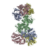 1e2tS S: Starting model for refinement |
|---|---|
| Similar structure data |

-Links
 PDBj
PDBj


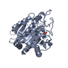
- Assembly
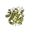
| Deposited unit | 
| ||||||||
|---|---|---|---|---|---|---|---|---|---|
| 1 |
| ||||||||
| Unit cell |
|
-Components
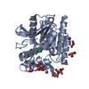
| #1: Protein | Mass: 35302.488 Da / Num. of mol.: 1 / Mutation: Y38F Source method: isolated from a genetically manipulated source Source: (gene. exp.) References: UniProt: Q81R98, UniProt: A0A6L7HKL6*PLUS, arylamine N-acetyltransferase | ||
|---|---|---|---|
| #2: Chemical | ChemComp-COA /   Mass: 767.534 Da / Num. of mol.: 1 / Source method: obtained synthetically / Formula: C21H36N7O16P3S Mass: 767.534 Da / Num. of mol.: 1 / Source method: obtained synthetically / Formula: C21H36N7O16P3S | ||
| #3: Chemical |   Mass: 46.025 Da / Num. of mol.: 2 / Source method: obtained synthetically / Formula: CH2O2 Mass: 46.025 Da / Num. of mol.: 2 / Source method: obtained synthetically / Formula: CH2O2#4: Water | ChemComp-HOH / |  Mass: 18.015 Da / Num. of mol.: 137 / Source method: isolated from a natural source / Formula: H2O Mass: 18.015 Da / Num. of mol.: 137 / Source method: isolated from a natural source / Formula: H2O |
-Experimental details
-Experiment
| Experiment | Method: X-RAY DIFFRACTION / Number of used crystals: 1 |
|---|
- Sample preparation
| Crystal | Density Matthews: 1.779778 Å3/Da / Density % sol: 30.890238 % |
|---|---|
| Crystal grow | Temperature: 294 K / Method: vapor diffusion Details: 1.8-1.9M Ammonium sulfate, 0.17M-0.2M potassium nitrate, VAPOR DIFFUSION, temperature 294K |
-Data collection
| Diffraction | Mean temperature: 100 K |
|---|---|
| Diffraction source | Source: SYNCHROTRON / Site: ESRF  / Beamline: ID23-1 / Wavelength: 0.92 Å / Beamline: ID23-1 / Wavelength: 0.92 Å |
| Detector | Type: ADSC QUANTUM 315r / Detector: CCD / Date: Nov 21, 2008 / Details: mirrors |
| Radiation | Monochromator: Silicon 1 1 1 channel / Protocol: SINGLE WAVELENGTH / Monochromatic (M) / Laue (L): M / Scattering type: x-ray |
| Radiation wavelength | Wavelength: 0.92 Å / Relative weight: 1 |
| Reflection | Resolution: 2→50 Å / Num. all: 18116 / Num. obs: 17034 / % possible obs: 94 % / Observed criterion σ(F): 0 / Observed criterion σ(I): -3 / Redundancy: 5.68 % / Biso Wilson estimate: 24.85 Å2 / Rmerge(I) obs: 0.062 / Rsym value: 0.063 / Net I/σ(I): 20.45 |
| Reflection shell | Resolution: 2→2.2 Å / Redundancy: 4.24 % / Rmerge(I) obs: 0.202 / Mean I/σ(I) obs: 7.91 / Num. unique all: 4366 / Rsym value: 0.177 / % possible all: 75.7 |
-Phasing
| Phasing | Method: molecular replacement | ||||||||||||||||||
|---|---|---|---|---|---|---|---|---|---|---|---|---|---|---|---|---|---|---|---|
| Phasing MR | Cor.coef. Fo:Fc: 0.69 / Packing: 0.682
|
- Processing
| Software |
| ||||||||||||||||||||||||||||||||||||
|---|---|---|---|---|---|---|---|---|---|---|---|---|---|---|---|---|---|---|---|---|---|---|---|---|---|---|---|---|---|---|---|---|---|---|---|---|---|
| Refinement | Method to determine structure: MOLECULAR REPLACEMENT Starting model: 1E2T Resolution: 2.01→45.76 Å / Rfactor Rfree error: 0.008 / Occupancy max: 1 / Occupancy min: 1 / Data cutoff high absF: 2205494 / Data cutoff low absF: 0 / Isotropic thermal model: RESTRAINED / Cross valid method: THROUGHOUT / σ(F): 0 / Stereochemistry target values: Engh & Huber / Details: BULK SOLVENT MODEL USED
| ||||||||||||||||||||||||||||||||||||
| Solvent computation | Solvent model: FLAT MODEL / Bsol: 39.02 Å2 / ksol: 0.4 e/Å3 | ||||||||||||||||||||||||||||||||||||
| Displacement parameters | Biso max: 44.33 Å2 / Biso mean: 19.19 Å2 / Biso min: 8.73 Å2
| ||||||||||||||||||||||||||||||||||||
| Refine analyze |
| ||||||||||||||||||||||||||||||||||||
| Refinement step | Cycle: LAST / Resolution: 2.01→45.76 Å
| ||||||||||||||||||||||||||||||||||||
| Refine LS restraints |
| ||||||||||||||||||||||||||||||||||||
| LS refinement shell | Resolution: 2.01→2.13 Å / Rfactor Rfree error: 0.022 / Total num. of bins used: 6
| ||||||||||||||||||||||||||||||||||||
| Xplor file |
|
