Resolution: 1.702→20 Å / Cor.coef. Fo:Fc: 0.941 / Cor.coef. Fo:Fc free: 0.918 / WRfactor Rfree: 0.219 / WRfactor Rwork: 0.182 / SU B: 2.029 / SU ML: 0.069 / Cross valid method: THROUGHOUT / σ(F): 0 / ESU R: 0.109 / ESU R Free: 0.11 / Stereochemistry target values: MAXIMUM LIKELIHOOD Details: HYDROGENS HAVE BEEN ADDED IN THE RIDING POSITIONS. U VALUES: REFINED INDIVIDUALLY. The programs coot and PRODRG and MOLPROBITY servers were also used during refinement.
Rfactor
Num. reflection
% reflection
Selection details
Rfree
0.231
2437
5.056 %
RANDOM
Rwork
0.191
-
-
-
obs
0.193
48202
98.714 %
-
Solvent computation
Ion probe radii: 0.8 Å / Shrinkage radii: 0.8 Å / VDW probe radii: 1.2 Å / Solvent model: MASK BULK SOLVENT
Displacement parameters
Biso mean: 13.169 Å2
Baniso -1
Baniso -2
Baniso -3
1-
-0.767 Å2
0 Å2
0 Å2
2-
-
0.176 Å2
0 Å2
3-
-
-
0.591 Å2
Refinement step
Cycle: LAST / Resolution: 1.702→20 Å
Protein
Nucleic acid
Ligand
Solvent
Total
Num. atoms
3036
0
53
294
3383
Refine LS restraints
Refine-ID
Type
Dev ideal
Dev ideal target
Number
X-RAY DIFFRACTION
r_bond_refined_d
0.016
0.022
3215
X-RAY DIFFRACTION
r_bond_other_d
0.001
0.02
2284
X-RAY DIFFRACTION
r_angle_refined_deg
1.474
1.957
4366
X-RAY DIFFRACTION
r_angle_other_deg
0.862
3.001
5453
X-RAY DIFFRACTION
r_dihedral_angle_1_deg
6.067
5
381
X-RAY DIFFRACTION
r_dihedral_angle_2_deg
28.748
22.289
166
X-RAY DIFFRACTION
r_dihedral_angle_3_deg
12.401
15
521
X-RAY DIFFRACTION
r_dihedral_angle_4_deg
12.148
15
33
X-RAY DIFFRACTION
r_chiral_restr
0.106
0.2
445
X-RAY DIFFRACTION
r_gen_planes_refined
0.007
0.021
3566
X-RAY DIFFRACTION
r_gen_planes_other
0.001
0.02
743
X-RAY DIFFRACTION
r_mcbond_it
2.414
2
1875
X-RAY DIFFRACTION
r_mcbond_other
0.816
2
753
X-RAY DIFFRACTION
r_mcangle_it
3.537
3
3009
X-RAY DIFFRACTION
r_scbond_it
3.058
2
1340
X-RAY DIFFRACTION
r_scangle_it
4.585
3
1352
LS refinement shell
Refine-ID: X-RAY DIFFRACTION / Total num. of bins used: 20
Resolution (Å)
Rfactor Rfree
Num. reflection Rfree
Rfactor Rwork
Num. reflection Rwork
Num. reflection all
% reflection obs (%)
1.702-1.746
0.321
177
0.253
3185
3526
95.349
1.746-1.793
0.281
181
0.21
3254
3473
98.906
1.793-1.845
0.233
158
0.182
3168
3345
99.432
1.845-1.901
0.24
150
0.193
3092
3261
99.417
1.901-1.963
0.215
165
0.196
2972
3157
99.366
1.963-2.031
0.258
144
0.186
2908
3070
99.414
2.031-2.107
0.249
158
0.192
2799
2967
99.663
2.107-2.192
0.207
166
0.187
2677
2857
99.51
2.192-2.288
0.24
133
0.187
2614
2761
99.493
2.288-2.398
0.189
136
0.183
2480
2628
99.543
2.398-2.526
0.234
136
0.185
2367
2513
99.602
2.526-2.677
0.23
110
0.191
2281
2400
99.625
2.677-2.858
0.229
121
0.193
2119
2250
99.556
2.858-3.081
0.251
97
0.21
1990
2100
99.381
3.081-3.368
0.252
95
0.209
1839
1950
99.179
3.368-3.752
0.228
85
0.192
1665
1785
98.039
3.752-4.307
0.184
77
0.163
1488
1597
97.996
4.307-5.215
0.187
73
0.158
1275
1371
98.322
5.215-7.136
0.29
48
0.197
1027
1101
97.639
7.136-20
0.259
27
0.23
565
718
82.451
+
About Yorodumi
-
News
-
Feb 9, 2022. New format data for meta-information of EMDB entries
New format data for meta-information of EMDB entries
Version 3 of the EMDB header file is now the official format.
The previous official version 1.9 will be removed from the archive.
In the structure databanks used in Yorodumi, some data are registered as the other names, "COVID-19 virus" and "2019-nCoV". Here are the details of the virus and the list of structure data.
Jan 31, 2019. EMDB accession codes are about to change! (news from PDBe EMDB page)
EMDB accession codes are about to change! (news from PDBe EMDB page)
The allocation of 4 digits for EMDB accession codes will soon come to an end. Whilst these codes will remain in use, new EMDB accession codes will include an additional digit and will expand incrementally as the available range of codes is exhausted. The current 4-digit format prefixed with “EMD-” (i.e. EMD-XXXX) will advance to a 5-digit format (i.e. EMD-XXXXX), and so on. It is currently estimated that the 4-digit codes will be depleted around Spring 2019, at which point the 5-digit format will come into force.
The EM Navigator/Yorodumi systems omit the EMD- prefix.
Related info.:Q: What is EMD? / ID/Accession-code notation in Yorodumi/EM Navigator
Yorodumi is a browser for structure data from EMDB, PDB, SASBDB, etc.
This page is also the successor to EM Navigator detail page, and also detail information page/front-end page for Omokage search.
The word "yorodu" (or yorozu) is an old Japanese word meaning "ten thousand". "mi" (miru) is to see.
Related info.:EMDB / PDB / SASBDB / Comparison of 3 databanks / Yorodumi Search / Aug 31, 2016. New EM Navigator & Yorodumi / Yorodumi Papers / Jmol/JSmol / Function and homology information / Changes in new EM Navigator and Yorodumi
 Movie
Movie Controller

 Yorodumi
 Open data
 Basic information
 Components
 Keywords
 Function and homology information
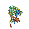
 Homo sapiens (human)
 X-RAY DIFFRACTION /
  Authors
 Citation
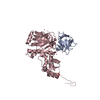
 Structure visualization
 Downloads & links
 Other downloads
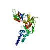
 PDBj
PDBj


 Assembly


 Mass: 65.409 Da / Num. of mol.: 1 / Source method: obtained synthetically / Formula: Zn
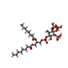
 Mass: 826.546 Da / Num. of mol.: 2 / Source method: obtained synthetically / Formula: C25H50O22P4
 Mass: 18.015 Da / Num. of mol.: 294 / Source method: isolated from a natural source / Formula: H2O
 Sample preparation
 / Beamline: 19-ID / Wavelength: 0.97918 Å
 Processing