 Movie
Movie Controller


+ Open data

- Basic information
| Entry | Database: PDB / ID: 3lje | ||||||
|---|---|---|---|---|---|---|---|
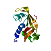
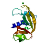
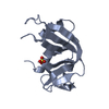
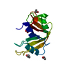
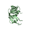
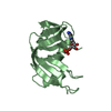
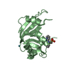
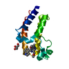
| Title | The X-ray structure of zebrafish RNase5 | ||||||
 Components Components | Zebrafish RNase5 | ||||||
 Keywords Keywords | HYDROLASE / angiogenins / fish rnases | ||||||
| Function / homology |  Function and homology information Function and homology informationendonuclease activity / nucleic acid binding / hydrolase activity / extracellular region Similarity search - Function | ||||||
| Biological species |  | ||||||
| Method |  X-RAY DIFFRACTION / MOLECULAR REPLACEMENT / Resolution: 1.8 Å X-RAY DIFFRACTION / MOLECULAR REPLACEMENT / Resolution: 1.8 Å | ||||||
 Authors Authors | Russo Krauss, I. / Merlino, A. / Coscia, F. / Mazzarella, L. / Sica, F. | ||||||
 Citation Citation | Journal: Biochem.J. / Year: 2010 Title: A new RNase sheds light on the RNase/angiogenin subfamily from zebrafish. Authors: Pizzo, E. / Merlino, A. / Turano, M. / Russo Krauss, I. / Coscia, F. / Zanfardino, A. / Varcamonti, M. / Furia, A. / Giancola, C. / Mazzarella, L. / Sica, F. / D'Alessio, G. #1: Journal: Gene / Year: 2007Title: The success of the RNase scaffold in the advance of bioscience and in evolution. Authors: Pizzo, E. / D'Alessio, G. #2: Journal: J.Biol.Chem. / Year: 2006Title: Ribonucleases and angiogenins from fish Authors: Pizzo, E. / Buonanno, P. / Di Maro, A. / Ponticelli, S. / De Falco, S. / Quarto, N. / Cubellis, M.V. / D'Alessio, G. | ||||||
| History |
|
- Structure visualization
| Structure viewer | Molecule: MolmilJmol/JSmol |
|---|
- Downloads & links
-Download
| PDBx/mmCIF format | 3lje.cif.gz | 40.7 KB | Display | PDBx/mmCIF format |
|---|---|---|---|---|
| PDB format | pdb3lje.ent.gz | 29.4 KB | Display | PDB format |
| PDBx/mmJSON format | 3lje.json.gz | Tree view | PDBx/mmJSON format | |
| Others |  Other downloads Other downloads |
-Validation report
| Arichive directory | https://data.pdbj.org/pub/pdb/validation_reports/lj/3ljeftp://data.pdbj.org/pub/pdb/validation_reports/lj/3lje | HTTPS FTP |
|---|
-Related structure data
| Related structure data | 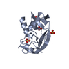 3ljdSC 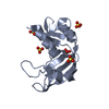 3ln8C S: Starting model for refinement C: citing same article ( |
|---|---|
| Similar structure data |


-Links
 PDBj
PDBj

- Assembly
| Deposited unit | 
| ||||||||
|---|---|---|---|---|---|---|---|---|---|
| 1 |
| ||||||||
| Unit cell |
|
-Components
| #1: Protein | Mass: 14533.582 Da / Num. of mol.: 1 Source method: isolated from a genetically manipulated source Source: (gene. exp.)  | ||||
|---|---|---|---|---|---|
| #2: Chemical | ChemComp-ACT /   Mass: 59.044 Da / Num. of mol.: 1 / Source method: obtained synthetically / Formula: C2H3O2 Mass: 59.044 Da / Num. of mol.: 1 / Source method: obtained synthetically / Formula: C2H3O2 | ||||
| #3: Chemical |   Mass: 96.063 Da / Num. of mol.: 2 / Source method: obtained synthetically / Formula: SO4 Mass: 96.063 Da / Num. of mol.: 2 / Source method: obtained synthetically / Formula: SO4#4: Water | ChemComp-HOH / |  Mass: 18.015 Da / Num. of mol.: 110 / Source method: isolated from a natural source / Formula: H2O Mass: 18.015 Da / Num. of mol.: 110 / Source method: isolated from a natural source / Formula: H2OHas protein modification | Y | |
-Experimental details
-Experiment
| Experiment | Method: X-RAY DIFFRACTION / Number of used crystals: 1 |
|---|
- Sample preparation
| Crystal | Density Matthews: 2.9 Å3/Da / Density % sol: 57.6 % |
|---|---|
| Crystal grow | Temperature: 293 K / pH: 4.5 Details: 32% MPEG 2K, 0.2M ammonium sulfate, 0.1M sodium acetate , pH 4.5, VAPOR DIFFUSION, HANGING DROP, temperature 293K |
-Data collection
| Diffraction | Mean temperature: 100 K |
|---|---|
| Diffraction source | Source: ROTATING ANODE / Type: RIGAKU / Wavelength: 1.5418 |
| Detector | Type: RIGAKU SATURN 944 / Detector: CCD / Date: Jul 27, 2009 / Details: MIRRORS |
| Radiation | Monochromator: GRAPHITE / Protocol: SINGLE WAVELENGTH / Monochromatic (M) / Laue (L): M / Scattering type: x-ray |
| Radiation wavelength | Wavelength: 1.5418 Å / Relative weight: 1 |
| Reflection | Resolution: 1.8→50 Å / Num. obs: 16327 / % possible obs: 99.9 % / Observed criterion σ(I): 0 / Rmerge(I) obs: 0.095 |
- Processing
| Software |
| |||||||||||||||||||||
|---|---|---|---|---|---|---|---|---|---|---|---|---|---|---|---|---|---|---|---|---|---|---|
| Refinement | Method to determine structure: MOLECULAR REPLACEMENT Starting model: 3LJD Resolution: 1.8→24.81 Å / Cross valid method: THROUGHOUT / σ(F): 0 / Stereochemistry target values: ENGH & HUBER
| |||||||||||||||||||||
| Refinement step | Cycle: LAST / Resolution: 1.8→24.81 Å
|
