 Movie
Movie Controller

[English] 日本語
 Yorodumi
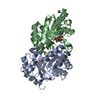
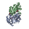
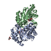
- PDB-3li1: Crystal structure of the mutant I218A of orotidine 5'-monophospha... -
+ Open data

- Basic information
| Entry | Database: PDB / ID: 3li1 | ||||||
|---|---|---|---|---|---|---|---|
| Title | Crystal structure of the mutant I218A of orotidine 5'-monophosphate decarboxylase from Methanobacterium thermoautotrophicum complexed with inhibitor BMP | ||||||
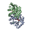
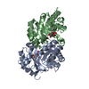
 Components Components | Orotidine 5'-phosphate decarboxylase | ||||||
 Keywords Keywords | LYASE / mutant I218A / 6-hydroxyuridine-5'-phosphate / Decarboxylase / Pyrimidine biosynthesis | ||||||
| Function / homology |  Function and homology information Function and homology informationorotidine-5'-phosphate decarboxylase / orotidine-5'-phosphate decarboxylase activity / 'de novo' UMP biosynthetic process / 'de novo' pyrimidine nucleobase biosynthetic process / cytosol Similarity search - Function | ||||||
| Biological species |   Methanothermobacter thermautotrophicus (archaea) Methanothermobacter thermautotrophicus (archaea) | ||||||
| Method |  X-RAY DIFFRACTION / SYNCHROTRON / MOLECULAR REPLACEMENT / Resolution: 1.35 Å X-RAY DIFFRACTION / SYNCHROTRON / MOLECULAR REPLACEMENT / Resolution: 1.35 Å | ||||||
 Authors Authors | Fedorov, A.A. / Fedorov, E.V. / Wood, B.M. / Gerlt, J.A. / Almo, S.C. | ||||||
 Citation Citation | Journal: Biochemistry / Year: 2010 Title: Conformational changes in orotidine 5'-monophosphate decarboxylase: "remote" residues that stabilize the active conformation. Authors: Wood, B.M. / Amyes, T.L. / Fedorov, A.A. / Fedorov, E.V. / Shabila, A. / Almo, S.C. / Richard, J.P. / Gerlt, J.A. | ||||||
| History |
|
- Structure visualization
| Structure viewer | Molecule: MolmilJmol/JSmol |
|---|
- Downloads & links
-Download
| PDBx/mmCIF format | 3li1.cif.gz | 100.2 KB | Display | PDBx/mmCIF format |
|---|---|---|---|---|
| PDB format | pdb3li1.ent.gz | 76 KB | Display | PDB format |
| PDBx/mmJSON format | 3li1.json.gz | Tree view | PDBx/mmJSON format | |
| Others |  Other downloads Other downloads |
-Validation report
| Arichive directory | https://data.pdbj.org/pub/pdb/validation_reports/li/3li1ftp://data.pdbj.org/pub/pdb/validation_reports/li/3li1 | HTTPS FTP |
|---|
-Related structure data
| Related structure data |  3lhtC 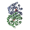 3lhuC 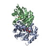 3lhvC  3lhwC 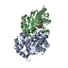 3lhyC  3lhzC 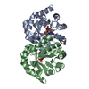 3lldC 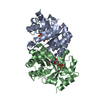 3llfC  3ltpC  3ltsC  3ltyC  3lv5C  3lv6C  3m1zC  3m41C  3m43C  3m44C  3m47C  3m5xC  3m5yC  3m5zC  3g18S C: citing same article ( S: Starting model for refinement |
|---|---|
| Similar structure data |


-Links
 PDBj
PDBj
- Assembly
| Deposited unit | 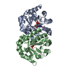
| ||||||||
|---|---|---|---|---|---|---|---|---|---|
| 1 |
| ||||||||
| Unit cell |
|
-Components
| #1: Protein | Mass: 24842.617 Da / Num. of mol.: 2 / Mutation: I218A Source method: isolated from a genetically manipulated source Source: (gene. exp.) Methanothermobacter thermautotrophicus (archaea)Strain: Delta H / Gene: pyrF, MTH_129 / Production host:  References: UniProt: O26232, orotidine-5'-phosphate decarboxylase #2: Chemical | 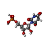  Type: RNA linking / Mass: 340.181 Da / Num. of mol.: 2 / Source method: obtained synthetically / Formula: C9H13N2O10P Type: RNA linking / Mass: 340.181 Da / Num. of mol.: 2 / Source method: obtained synthetically / Formula: C9H13N2O10P#3: Water | ChemComp-HOH / |  Mass: 18.015 Da / Num. of mol.: 282 / Source method: isolated from a natural source / Formula: H2O Mass: 18.015 Da / Num. of mol.: 282 / Source method: isolated from a natural source / Formula: H2O |
|---|
-Experimental details
-Experiment
| Experiment | Method: X-RAY DIFFRACTION / Number of used crystals: 1 |
|---|
- Sample preparation
| Crystal | Density Matthews: 2.12 Å3/Da / Density % sol: 41.98 % |
|---|---|
| Crystal grow | Temperature: 293 K / Method: vapor diffusion, hanging drop / pH: 7.5 Details: 1.4M sodium citrate, 0.1M Hepes, pH 7.5, VAPOR DIFFUSION, HANGING DROP, temperature 293.0K |
-Data collection
| Diffraction | Mean temperature: 100 K |
|---|---|
| Diffraction source | Source: SYNCHROTRON / Site: NSLS  / Beamline: X4A / Wavelength: 0.97915 Å / Beamline: X4A / Wavelength: 0.97915 Å |
| Detector | Type: ADSC QUANTUM 4 / Detector: CCD / Date: Nov 11, 2009 |
| Radiation | Monochromator: Si 111 CHANNEL / Protocol: SINGLE WAVELENGTH / Monochromatic (M) / Laue (L): M / Scattering type: x-ray |
| Radiation wavelength | Wavelength: 0.97915 Å / Relative weight: 1 |
| Reflection | Resolution: 1.35→25 Å / Num. all: 85377 / Num. obs: 85377 / % possible obs: 93.6 % / Observed criterion σ(F): 0 / Observed criterion σ(I): 0 / Biso Wilson estimate: 16 Å2 / Rmerge(I) obs: 0.054 |
- Processing
| Software |
| ||||||||||||||||||||||||||||||||||||
|---|---|---|---|---|---|---|---|---|---|---|---|---|---|---|---|---|---|---|---|---|---|---|---|---|---|---|---|---|---|---|---|---|---|---|---|---|---|
| Refinement | Method to determine structure: MOLECULAR REPLACEMENT Starting model: 3G18 Resolution: 1.35→22.72 Å / Rfactor Rfree error: 0.003 / Data cutoff high absF: 1081617.67 / Data cutoff low absF: 0 / Isotropic thermal model: RESTRAINED / Cross valid method: THROUGHOUT / σ(F): 0 / σ(I): 0 / Stereochemistry target values: Engh & Huber
| ||||||||||||||||||||||||||||||||||||
| Solvent computation | Solvent model: FLAT MODEL / Bsol: 55.432 Å2 / ksol: 0.410797 e/Å3 | ||||||||||||||||||||||||||||||||||||
| Displacement parameters | Biso mean: 18.2 Å2
| ||||||||||||||||||||||||||||||||||||
| Refine analyze |
| ||||||||||||||||||||||||||||||||||||
| Refinement step | Cycle: LAST / Resolution: 1.35→22.72 Å
| ||||||||||||||||||||||||||||||||||||
| Refine LS restraints |
| ||||||||||||||||||||||||||||||||||||
| LS refinement shell | Resolution: 1.35→1.4 Å / Rfactor Rfree error: 0.017 / Total num. of bins used: 10
| ||||||||||||||||||||||||||||||||||||
| Xplor file |
|
