 Movie
Movie Controller


+ Open data

- Basic information
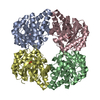
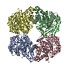
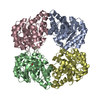
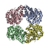
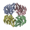
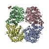
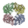
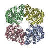
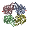
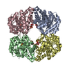
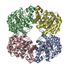
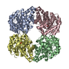
| Entry | Database: PDB / ID: 3lch | ||||||
|---|---|---|---|---|---|---|---|
| Title | The D-sialic acid aldolase mutant V251R | ||||||
 Components Components | N-acetylneuraminate lyase | ||||||
 Keywords Keywords | LYASE / Tim barrel / Carbohydrate metabolism / Schiff base | ||||||
| Function / homology |  Function and homology information Function and homology informationN-acetylneuraminate lyase / N-acetylneuraminate lyase activity / N-acetylneuraminate catabolic process / single-species biofilm formation / carbohydrate metabolic process / identical protein binding / cytosol Similarity search - Function | ||||||
| Biological species |  | ||||||
| Method |  X-RAY DIFFRACTION / SYNCHROTRON / MOLECULAR REPLACEMENT / Resolution: 2.04 Å X-RAY DIFFRACTION / SYNCHROTRON / MOLECULAR REPLACEMENT / Resolution: 2.04 Å | ||||||
 Authors Authors | Chou, C.-Y. / Wang, A.H.-J. / Ko, T.-P. | ||||||
 Citation Citation | Journal: To be Published Title: Modulation of substrate specificities of D-sialic acid aldolase through single mutations of Val251 Authors: Chou, C.-Y. / Ko, T.-P. / Wu, K.-J. / Huang, K.-F. / Lin, C.-H. / Wong, C.-H. / Wang, A.H.-J. | ||||||
| History |
|
- Structure visualization
| Structure viewer | Molecule: MolmilJmol/JSmol |
|---|
- Downloads & links
-Download
| PDBx/mmCIF format | 3lch.cif.gz | 252.3 KB | Display | PDBx/mmCIF format |
|---|---|---|---|---|
| PDB format | pdb3lch.ent.gz | 201.2 KB | Display | PDB format |
| PDBx/mmJSON format | 3lch.json.gz | Tree view | PDBx/mmJSON format | |
| Others |  Other downloads Other downloads |
-Validation report
| Arichive directory | https://data.pdbj.org/pub/pdb/validation_reports/lc/3lchftp://data.pdbj.org/pub/pdb/validation_reports/lc/3lch | HTTPS FTP |
|---|
-Related structure data
| Related structure data |  3lbcC  3lbmC  3lcfC  3lcgC  3lciC  3lclC  3lcwC  3lcxC  1nalS S: Starting model for refinement C: citing same article ( |
|---|---|
| Similar structure data |


-Links
 PDBj
PDBj- Assembly
| Deposited unit | 
| ||||||||
|---|---|---|---|---|---|---|---|---|---|
| 1 |
| ||||||||
| Unit cell |
|
-Components
| #1: Protein | Mass: 35349.355 Da / Num. of mol.: 4 / Mutation: V251R Source method: isolated from a genetically manipulated source Source: (gene. exp.) #2: Chemical | ChemComp-SO4 /   Mass: 96.063 Da / Num. of mol.: 4 / Source method: obtained synthetically / Formula: SO4 Mass: 96.063 Da / Num. of mol.: 4 / Source method: obtained synthetically / Formula: SO4#3: Water | ChemComp-HOH / |  Mass: 18.015 Da / Num. of mol.: 891 / Source method: isolated from a natural source / Formula: H2O Mass: 18.015 Da / Num. of mol.: 891 / Source method: isolated from a natural source / Formula: H2O |
|---|
-Experimental details
-Experiment
| Experiment | Method: X-RAY DIFFRACTION / Number of used crystals: 1 |
|---|
- Sample preparation
| Crystal | Density Matthews: 3.03 Å3/Da / Density % sol: 59.39 % |
|---|---|
| Crystal grow | Temperature: 298 K / Method: vapor diffusion, hanging drop / pH: 6.5 Details: 2.0M ammonium sulfate, 0.1M Bis-Tris, pH 6.5, VAPOR DIFFUSION, HANGING DROP, temperature 298K |
-Data collection
| Diffraction | Mean temperature: 100 K |
|---|---|
| Diffraction source | Source: SYNCHROTRON / Site: NSRRC  / Beamline: BL13C1 / Wavelength: 0.97622 Å / Beamline: BL13C1 / Wavelength: 0.97622 Å |
| Detector | Type: ADSC QUANTUM 210 / Detector: CCD / Date: Apr 23, 2009 / Details: vertically focusing mirror |
| Radiation | Monochromator: horizontally focusing single crystal Si(111) bent Monochromator Protocol: SINGLE WAVELENGTH / Monochromatic (M) / Laue (L): M / Scattering type: x-ray |
| Radiation wavelength | Wavelength: 0.97622 Å / Relative weight: 1 |
| Reflection | Resolution: 2.04→30 Å / Num. all: 109845 / Num. obs: 109625 / % possible obs: 99.8 % / Observed criterion σ(F): 0 / Observed criterion σ(I): 1 / Redundancy: 6.1 % / Rmerge(I) obs: 0.074 / Net I/σ(I): 30.4 |
| Reflection shell | Resolution: 2.04→2.11 Å / Redundancy: 6.1 % / Rmerge(I) obs: 0.458 / Mean I/σ(I) obs: 3.7 / Num. unique all: 10837 / % possible all: 100 |
- Processing
| Software |
| ||||||||||||||||||||||||||||
|---|---|---|---|---|---|---|---|---|---|---|---|---|---|---|---|---|---|---|---|---|---|---|---|---|---|---|---|---|---|
| Refinement | Method to determine structure: MOLECULAR REPLACEMENT Starting model: PDB enrty 1NAL Resolution: 2.04→28.2 Å / Cross valid method: THROUGHOUT / σ(F): 0 / σ(I): 0 / Stereochemistry target values: Engh & Huber
| ||||||||||||||||||||||||||||
| Solvent computation | Bsol: 63.968 Å2 | ||||||||||||||||||||||||||||
| Displacement parameters | Biso mean: 32.298 Å2
| ||||||||||||||||||||||||||||
| Refine analyze |
| ||||||||||||||||||||||||||||
| Refinement step | Cycle: LAST / Resolution: 2.04→28.2 Å
| ||||||||||||||||||||||||||||
| Refine LS restraints |
| ||||||||||||||||||||||||||||
| LS refinement shell | Resolution: 2.04→2.11 Å / Rfactor Rfree error: 0.0241
| ||||||||||||||||||||||||||||
| Xplor file |
|
