 Movie
Movie Controller


+ Open data

- Basic information
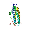
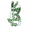
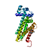
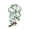
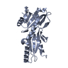
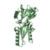
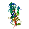
| Entry | Database: PDB / ID: 3l9t | ||||||
|---|---|---|---|---|---|---|---|
| Title | The Crystal Structure of smu.31 from Streptococcus mutans UA159 | ||||||
 Components Components | Putative uncharacterized protein smu.31 | ||||||
 Keywords Keywords | UNKNOWN FUNCTION / hypothetical protein | ||||||
| Function / homology |  Function and homology information Function and homology informationMethyltransferase, Methionine Synthase (B12-binding Domains); Chain A, domain 1 - #70 / ARM repeat domains / DNA alkylation repair enzyme / DNA alkylation repair enzyme / Methyltransferase, Methionine Synthase (B12-binding Domains); Chain A, domain 1 / Serine Threonine Protein Phosphatase 5, Tetratricopeptide repeat / Alpha Horseshoe / Armadillo-type fold / Orthogonal Bundle / Mainly Alpha Similarity search - Domain/homology | ||||||
| Biological species |  Streptococcus mutans (bacteria) Streptococcus mutans (bacteria) | ||||||
| Method |  X-RAY DIFFRACTION / SYNCHROTRON / SAD / Resolution: 2.206 Å X-RAY DIFFRACTION / SYNCHROTRON / SAD / Resolution: 2.206 Å | ||||||
 Authors Authors | Su, X.-D. / Cao, Q. / Liu, X. | ||||||
 Citation Citation | Journal: TO BE PUBLISHED Title: The Crystal Structure of smu.31 from Streptococcus mutans UA159 Authors: Su, X.-D. / Cao, Q. / Liu, X. | ||||||
| History |
|
- Structure visualization
| Structure viewer | Molecule: MolmilJmol/JSmol |
|---|
- Downloads & links
-Download
| PDBx/mmCIF format | 3l9t.cif.gz | 50.9 KB | Display | PDBx/mmCIF format |
|---|---|---|---|---|
| PDB format | pdb3l9t.ent.gz | 39.4 KB | Display | PDB format |
| PDBx/mmJSON format | 3l9t.json.gz | Tree view | PDBx/mmJSON format | |
| Others |  Other downloads Other downloads |
-Validation report
| Summary document | 3l9t_validation.pdf.gz | 437.5 KB | Display | wwPDB validaton report |
|---|---|---|---|---|
| Full document | 3l9t_full_validation.pdf.gz | 439.7 KB | Display | |
| Data in XML | 3l9t_validation.xml.gz | 9.7 KB | Display | |
| Data in CIF | 3l9t_validation.cif.gz | 12.4 KB | Display | |
| Arichive directory | https://data.pdbj.org/pub/pdb/validation_reports/l9/3l9tftp://data.pdbj.org/pub/pdb/validation_reports/l9/3l9t | HTTPS FTP |
-Related structure data
| Similar structure data |
|---|


-Links
 PDBj
PDBj- Assembly
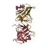
| Deposited unit | 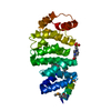
| ||||||||
|---|---|---|---|---|---|---|---|---|---|
| 1 |
| ||||||||
| Unit cell |
|
-Components
| #1: Protein | Mass: 28449.469 Da / Num. of mol.: 1 Source method: isolated from a genetically manipulated source Source: (gene. exp.) Streptococcus mutans (bacteria) / Strain: UA159 / Gene: smu.31 / Plasmid: pET28a / Production host: |
|---|---|
| #2: Chemical | ChemComp-EPE /   Mass: 238.305 Da / Num. of mol.: 1 / Source method: obtained synthetically / Formula: C8H18N2O4S / Comment: pH buffer*YM Mass: 238.305 Da / Num. of mol.: 1 / Source method: obtained synthetically / Formula: C8H18N2O4S / Comment: pH buffer*YM |
| #3: Water | ChemComp-HOH /  Mass: 18.015 Da / Num. of mol.: 14 / Source method: isolated from a natural source / Formula: H2O Mass: 18.015 Da / Num. of mol.: 14 / Source method: isolated from a natural source / Formula: H2O |
-Experimental details
-Experiment
| Experiment | Method: X-RAY DIFFRACTION / Number of used crystals: 1 |
|---|
- Sample preparation
| Crystal | Density Matthews: 2.1 Å3/Da / Density % sol: 41.38 % |
|---|---|
| Crystal grow | Temperature: 289 K / Method: vapor diffusion, sitting drop / pH: 7.5 Details: 14% PEG 6000, 0.1M HEPES pH7.5, VAPOR DIFFUSION, SITTING DROP, temperature 289K |
-Data collection
| Diffraction | Mean temperature: 100 K |
|---|---|
| Diffraction source | Source: SYNCHROTRON / Site: Photon Factory  / Beamline: BL-17A / Wavelength: 0.9789 Å / Beamline: BL-17A / Wavelength: 0.9789 Å |
| Detector | Type: ADSC QUANTUM 270 / Detector: CCD / Date: Jun 1, 2009 |
| Radiation | Protocol: SINGLE WAVELENGTH / Monochromatic (M) / Laue (L): M / Scattering type: x-ray |
| Radiation wavelength | Wavelength: 0.9789 Å / Relative weight: 1 |
| Reflection | Resolution: 2.2→50 Å / Num. all: 12788 / Num. obs: 12558 / % possible obs: 98.2 % / Observed criterion σ(I): 1 / Biso Wilson estimate: 23.98 Å2 |
| Reflection shell | Resolution: 2.2→2.3 Å / % possible all: 86.7 |
- Processing
| Software |
| ||||||||||||||||||||||||||||||||||||||||||||||||||||||||||||||||||||||
|---|---|---|---|---|---|---|---|---|---|---|---|---|---|---|---|---|---|---|---|---|---|---|---|---|---|---|---|---|---|---|---|---|---|---|---|---|---|---|---|---|---|---|---|---|---|---|---|---|---|---|---|---|---|---|---|---|---|---|---|---|---|---|---|---|---|---|---|---|---|---|---|
| Refinement | Method to determine structure: SAD / Resolution: 2.206→19.569 Å / Occupancy max: 1 / Occupancy min: 0.8 / FOM work R set: 0.836 / SU ML: 0.32 / σ(F): 2.03 / Phase error: 22.72 / Stereochemistry target values: ML
| ||||||||||||||||||||||||||||||||||||||||||||||||||||||||||||||||||||||
| Solvent computation | Shrinkage radii: 0.9 Å / VDW probe radii: 1.11 Å / Solvent model: FLAT BULK SOLVENT MODEL / Bsol: 48.907 Å2 / ksol: 0.454 e/Å3 | ||||||||||||||||||||||||||||||||||||||||||||||||||||||||||||||||||||||
| Displacement parameters | Biso max: 64.01 Å2 / Biso mean: 24.455 Å2 / Biso min: 9.6 Å2
| ||||||||||||||||||||||||||||||||||||||||||||||||||||||||||||||||||||||
| Refinement step | Cycle: LAST / Resolution: 2.206→19.569 Å
| ||||||||||||||||||||||||||||||||||||||||||||||||||||||||||||||||||||||
| Refine LS restraints |
| ||||||||||||||||||||||||||||||||||||||||||||||||||||||||||||||||||||||
| LS refinement shell |
|
