 Movie
Movie Controller


+ Open data

- Basic information
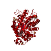
| Entry | Database: PDB / ID: 3l66 | ||||||
|---|---|---|---|---|---|---|---|
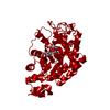
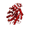
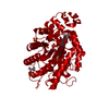
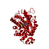
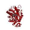
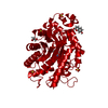
| Title | Xenobiotic Reductase A - C25A Variant with Coumarin | ||||||
 Components Components | Xenobiotic reductase A | ||||||
 Keywords Keywords | OXIDOREDUCTASE / FMN / flavin / old-yellow-enzyme | ||||||
| Function / homology |  Function and homology information Function and homology information | ||||||
| Biological species |  Pseudomonas putida (bacteria) Pseudomonas putida (bacteria) | ||||||
| Method |  X-RAY DIFFRACTION / SYNCHROTRON / FOURIER SYNTHESIS / Resolution: 1.28 Å X-RAY DIFFRACTION / SYNCHROTRON / FOURIER SYNTHESIS / Resolution: 1.28 Å | ||||||
 Authors Authors | Spiegelhauer, O. / Dobbek, H. | ||||||
 Citation Citation | Journal: J.Mol.Biol. / Year: 2010 Title: Cysteine as a modulator residue in the active site of xenobiotic reductase A: a structural, thermodynamic and kinetic study Authors: Spiegelhauer, O. / Mende, S. / Dickert, F. / Knauer, S.H. / Ullmann, G.M. / Dobbek, H. | ||||||
| History |
|
- Structure visualization
| Structure viewer | Molecule: MolmilJmol/JSmol |
|---|
- Downloads & links
-Download
| PDBx/mmCIF format | 3l66.cif.gz | 327 KB | Display | PDBx/mmCIF format |
|---|---|---|---|---|
| PDB format | pdb3l66.ent.gz | 264.6 KB | Display | PDB format |
| PDBx/mmJSON format | 3l66.json.gz | Tree view | PDBx/mmJSON format | |
| Others |  Other downloads Other downloads |
-Validation report
| Arichive directory | https://data.pdbj.org/pub/pdb/validation_reports/l6/3l66ftp://data.pdbj.org/pub/pdb/validation_reports/l6/3l66 | HTTPS FTP |
|---|
-Related structure data
| Related structure data |  3l5lC  3l5mC 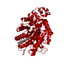 3l65SC  3l67C 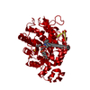 3l68C C: citing same article ( S: Starting model for refinement |
|---|---|
| Similar structure data |


-Links
 PDBj
PDBj- Assembly
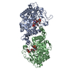
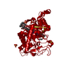
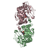
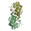
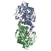
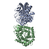
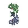
| Deposited unit | 
| ||||||||
|---|---|---|---|---|---|---|---|---|---|
| 1 | 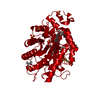
| ||||||||
| Unit cell |
| ||||||||
| Components on special symmetry positions |
|
-Components
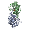
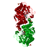
-Protein , 1 types, 1 molecules A
| #1: Protein | Mass: 39881.953 Da / Num. of mol.: 1 / Mutation: C25A Source method: isolated from a genetically manipulated source Source: (gene. exp.) Pseudomonas putida (bacteria) / Strain: 86 / Gene: xenA / Plasmid: pET / Production host: |
|---|
-Non-polymers , 5 types, 643 molecules 


| #2: Chemical | ChemComp-SO4 /  Mass: 96.063 Da / Num. of mol.: 6 / Source method: obtained synthetically / Formula: SO4 Mass: 96.063 Da / Num. of mol.: 6 / Source method: obtained synthetically / Formula: SO4#3: Chemical |  Mass: 90.121 Da / Num. of mol.: 2 / Source method: obtained synthetically / Formula: C4H10O2 Mass: 90.121 Da / Num. of mol.: 2 / Source method: obtained synthetically / Formula: C4H10O2#4: Chemical |  Mass: 146.143 Da / Num. of mol.: 3 / Source method: obtained synthetically / Formula: C9H6O2 Mass: 146.143 Da / Num. of mol.: 3 / Source method: obtained synthetically / Formula: C9H6O2#5: Chemical | ChemComp-FMN / |  Mass: 456.344 Da / Num. of mol.: 1 / Source method: obtained synthetically / Formula: C17H21N4O9P Mass: 456.344 Da / Num. of mol.: 1 / Source method: obtained synthetically / Formula: C17H21N4O9P#6: Water | ChemComp-HOH / | Mass: 18.015 Da / Num. of mol.: 631 / Source method: isolated from a natural source / Formula: H2O |
|---|
-Details
| Sequence details | A SEQUENCE DATABASE REFERENCE WHICH DERIVES FROM PSEUDOMONAS PUTIDA STRAIN 86 DOES NOT CURRENTLY ...A SEQUENCE DATABASE REFERENCE WHICH DERIVES FROM PSEUDOMONA |
|---|
-Experimental details
-Experiment
| Experiment | Method: X-RAY DIFFRACTION / Number of used crystals: 1 |
|---|
- Sample preparation
| Crystal | Density Matthews: 2.38 Å3/Da / Density % sol: 48.31 % |
|---|---|
| Crystal grow | Temperature: 290 K / Method: vapor diffusion, hanging drop / pH: 7.5 Details: 1.5M Ammonium sulfate, 100mM Hepes, pH 7.5, VAPOR DIFFUSION, HANGING DROP, temperature 290K |
-Data collection
| Diffraction | Mean temperature: 100 K |
|---|---|
| Diffraction source | Source: SYNCHROTRON / Site: BESSY  / Beamline: 14.2 / Wavelength: 0.92 Å / Beamline: 14.2 / Wavelength: 0.92 Å |
| Detector | Type: RAYONIX MX-225 / Detector: CCD |
| Radiation | Protocol: SINGLE WAVELENGTH / Monochromatic (M) / Laue (L): M / Scattering type: x-ray |
| Radiation wavelength | Wavelength: 0.92 Å / Relative weight: 1 |
| Reflection | Resolution: 1.28→33.11 Å / Num. all: 98065 / Num. obs: 96595 / % possible obs: 98.5 % / Observed criterion σ(F): 0 / Observed criterion σ(I): 0 / Biso Wilson estimate: 8.88 Å2 |
- Processing
| Software |
| |||||||||||||||||||||||||||||||||||||||||||||||||||||||||||||||||||||||||||||
|---|---|---|---|---|---|---|---|---|---|---|---|---|---|---|---|---|---|---|---|---|---|---|---|---|---|---|---|---|---|---|---|---|---|---|---|---|---|---|---|---|---|---|---|---|---|---|---|---|---|---|---|---|---|---|---|---|---|---|---|---|---|---|---|---|---|---|---|---|---|---|---|---|---|---|---|---|---|---|
| Refinement | Method to determine structure: FOURIER SYNTHESIS Starting model: 3L65 Resolution: 1.28→33.11 Å / Occupancy max: 1 / Occupancy min: 0 / FOM work R set: 0.915 / SU ML: 0.13 / Cross valid method: THROUGHOUT / σ(F): 1.99 / Phase error: 15 / Stereochemistry target values: ML
| |||||||||||||||||||||||||||||||||||||||||||||||||||||||||||||||||||||||||||||
| Solvent computation | Shrinkage radii: 0.9 Å / VDW probe radii: 1.11 Å / Solvent model: FLAT BULK SOLVENT MODEL / Bsol: 58.885 Å2 / ksol: 0.36 e/Å3 | |||||||||||||||||||||||||||||||||||||||||||||||||||||||||||||||||||||||||||||
| Displacement parameters | Biso max: 420.95 Å2 / Biso mean: 18.878 Å2 / Biso min: 4.88 Å2
| |||||||||||||||||||||||||||||||||||||||||||||||||||||||||||||||||||||||||||||
| Refinement step | Cycle: LAST / Resolution: 1.28→33.11 Å
| |||||||||||||||||||||||||||||||||||||||||||||||||||||||||||||||||||||||||||||
| Refine LS restraints |
| |||||||||||||||||||||||||||||||||||||||||||||||||||||||||||||||||||||||||||||
| LS refinement shell | Refine-ID: X-RAY DIFFRACTION / Total num. of bins used: 10
|
