 Movie
Movie Controller


+ Open data

- Basic information
| Entry | Database: PDB / ID: 3l5x | |||||||||
|---|---|---|---|---|---|---|---|---|---|---|
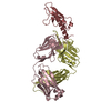
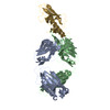
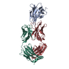
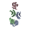
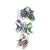
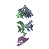
| Title | Crystal structure of the complex between IL-13 and H2L6 FAB | |||||||||
 Components Components |
| |||||||||
 Keywords Keywords | IMMUNE SYSTEM / immunoglobulin fold / alpha-helical bundle / Cytokine / Disulfide bond / Glycoprotein / Polymorphism / Secreted / MONOCLONAL ANTIBODY | |||||||||
| Function / homology |  Function and homology information Function and homology informationinterleukin-13 receptor binding / negative regulation of lung ciliated cell differentiation / positive regulation of lung goblet cell differentiation / positive regulation of pancreatic stellate cell proliferation / interleukin-13-mediated signaling pathway / regulation of proton transport / negative regulation of complement-dependent cytotoxicity / Interleukin-18 signaling / negative regulation of transforming growth factor beta production / positive regulation of mast cell degranulation ...interleukin-13 receptor binding / negative regulation of lung ciliated cell differentiation / positive regulation of lung goblet cell differentiation / positive regulation of pancreatic stellate cell proliferation / interleukin-13-mediated signaling pathway / regulation of proton transport / negative regulation of complement-dependent cytotoxicity / Interleukin-18 signaling / negative regulation of transforming growth factor beta production / positive regulation of mast cell degranulation / response to selenium ion / macrophage activation / response to nematode / positive regulation of macrophage activation / positive regulation of immunoglobulin production / positive regulation of interleukin-10 production / negative regulation of endothelial cell apoptotic process / positive regulation of B cell proliferation / positive regulation of smooth muscle cell proliferation / cytokine activity / positive regulation of release of sequestered calcium ion into cytosol / positive regulation of protein secretion / response to nicotine / cellular response to mechanical stimulus / microglial cell activation / negative regulation of inflammatory response / positive regulation of cold-induced thermogenesis / Interleukin-4 and Interleukin-13 signaling / response to lipopolysaccharide / response to ethanol / immune response / inflammatory response / external side of plasma membrane / positive regulation of gene expression / positive regulation of transcription by RNA polymerase II / : / extracellular region / cytoplasm Similarity search - Function | |||||||||
| Biological species |  Homo sapiens (human) Homo sapiens (human) | |||||||||
| Method |  X-RAY DIFFRACTION / MOLECULAR REPLACEMENT / Resolution: 1.9 Å X-RAY DIFFRACTION / MOLECULAR REPLACEMENT / Resolution: 1.9 Å | |||||||||
 Authors Authors | Teplyakov, A. / Obmolova, G. / Malia, T. / Gilliland, G.L. | |||||||||
 Citation Citation | Journal: J.Mol.Biol. / Year: 2010 Title: Human framework adaptation of a mouse anti-human IL-13 antibody. Authors: Fransson, J. / Teplyakov, A. / Raghunathan, G. / Chi, E. / Cordier, W. / Dinh, T. / Feng, Y. / Giles-Komar, J. / Gilliland, G. / Lollo, B. / Malia, T.J. / Nishioka, W. / Obmolova, G. / Zhao, ...Authors: Fransson, J. / Teplyakov, A. / Raghunathan, G. / Chi, E. / Cordier, W. / Dinh, T. / Feng, Y. / Giles-Komar, J. / Gilliland, G. / Lollo, B. / Malia, T.J. / Nishioka, W. / Obmolova, G. / Zhao, S. / Zhao, Y. / Swanson, R.V. / Almagro, J.C. | |||||||||
| History |
|
- Structure visualization
| Structure viewer | Molecule: MolmilJmol/JSmol |
|---|
- Downloads & links
-Download
| PDBx/mmCIF format | 3l5x.cif.gz | 126.7 KB | Display | PDBx/mmCIF format |
|---|---|---|---|---|
| PDB format | pdb3l5x.ent.gz | 95.7 KB | Display | PDB format |
| PDBx/mmJSON format | 3l5x.json.gz | Tree view | PDBx/mmJSON format | |
| Others |  Other downloads Other downloads |
-Validation report
| Arichive directory | https://data.pdbj.org/pub/pdb/validation_reports/l5/3l5xftp://data.pdbj.org/pub/pdb/validation_reports/l5/3l5x | HTTPS FTP |
|---|
-Related structure data
| Related structure data | 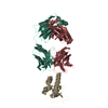 3l5wC  3l7fC 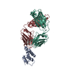 4ps4C  1q9q C: citing same article ( S: Starting model for refinement |
|---|---|
| Similar structure data |

-Links
 PDBj
PDBj


- Assembly
| Deposited unit | 
| ||||||||
|---|---|---|---|---|---|---|---|---|---|
| 1 |
| ||||||||
| Unit cell |
|
-Components
-Protein , 1 types, 1 molecules A
| #3: Protein | Mass: 12490.583 Da / Num. of mol.: 1 / Fragment: UNP residues 35-146 Source method: isolated from a genetically manipulated source Source: (gene. exp.) Homo sapiens (human) / Gene: IL13, NC30 / Production host:  |
|---|
-Antibody , 2 types, 2 molecules LH
| #1: Antibody | Mass: 23487.115 Da / Num. of mol.: 1 Source method: isolated from a genetically manipulated source Source: (gene. exp.) Homo sapiens (human) / Cell line (production host): HEK cells / Production host: HOMO SAPIENS (human) |
|---|---|
| #2: Antibody | Mass: 24110.328 Da / Num. of mol.: 1 Source method: isolated from a genetically manipulated source Source: (gene. exp.) Homo sapiens (human) / Cell line (production host): HEK cells / Production host: HOMO SAPIENS (human) |
-Non-polymers , 3 types, 354 molecules 


| #4: Chemical | ChemComp-MES /  Mass: 195.237 Da / Num. of mol.: 1 / Source method: obtained synthetically / Formula: C6H13NO4S / Comment: pH buffer*YM Mass: 195.237 Da / Num. of mol.: 1 / Source method: obtained synthetically / Formula: C6H13NO4S / Comment: pH buffer*YM |
|---|---|
| #5: Chemical | ChemComp-GOL /  Mass: 92.094 Da / Num. of mol.: 1 / Source method: obtained synthetically / Formula: C3H8O3 Mass: 92.094 Da / Num. of mol.: 1 / Source method: obtained synthetically / Formula: C3H8O3 |
| #6: Water | ChemComp-HOH / Mass: 18.015 Da / Num. of mol.: 352 / Source method: isolated from a natural source / Formula: H2O |
-Details
| Has protein modification | Y |
|---|
-Experimental details
-Experiment
| Experiment | Method: X-RAY DIFFRACTION / Number of used crystals: 1 |
|---|
- Sample preparation
| Crystal | Density Matthews: 2.23 Å3/Da / Density % sol: 45 % |
|---|---|
| Crystal grow | Temperature: 293 K / Method: vapor diffusion, sitting drop / pH: 6.5 Details: 0.1 M MES PH 6.5, 14% PEG 3350, 0.2 M AMMONIUM TARTRATE. CRYO CONDITIONS: 0.1 M MES PH 6.5, 20% PEG 3K, 0.2 M AMM TARTRATE, 15% GLYCEROL, VAPOR DIFFUSION, SITTING DROP, temperature 293K |
-Data collection
| Diffraction | Mean temperature: 100 K |
|---|---|
| Diffraction source | Source: ROTATING ANODE / Type: RIGAKU MICROMAX-007 HF / Wavelength: 1.5418 / Wavelength: 1.5418 Å |
| Detector | Type: RIGAKU SATURN 944 / Detector: CCD / Date: Jan 15, 2008 / Details: VARIMAX HF |
| Radiation | Protocol: SINGLE WAVELENGTH / Monochromatic (M) / Laue (L): M / Scattering type: x-ray |
| Radiation wavelength | Wavelength: 1.5418 Å / Relative weight: 1 |
| Reflection | Resolution: 1.9→64 Å / Num. all: 42621 / Num. obs: 42621 / % possible obs: 98.9 % / Observed criterion σ(I): -3 / Redundancy: 16.3 % / Biso Wilson estimate: 36.5 Å2 / Rmerge(I) obs: 0.084 / Net I/σ(I): 14.5 |
| Reflection shell | Resolution: 1.9→1.97 Å / Redundancy: 3.8 % / Rmerge(I) obs: 0.392 / Mean I/σ(I) obs: 2.3 / % possible all: 91.1 |
- Processing
| Software |
| ||||||||||||||||||||||||||||||||||||||||||||||||||||||||||||||||||||||||||||||||||||||||||
|---|---|---|---|---|---|---|---|---|---|---|---|---|---|---|---|---|---|---|---|---|---|---|---|---|---|---|---|---|---|---|---|---|---|---|---|---|---|---|---|---|---|---|---|---|---|---|---|---|---|---|---|---|---|---|---|---|---|---|---|---|---|---|---|---|---|---|---|---|---|---|---|---|---|---|---|---|---|---|---|---|---|---|---|---|---|---|---|---|---|---|---|
| Refinement | Method to determine structure: MOLECULAR REPLACEMENT Starting model: PDB ENTRY 1Q9Q 1q9q Resolution: 1.9→15 Å / Cor.coef. Fo:Fc: 0.961 / Cor.coef. Fo:Fc free: 0.946 / SU B: 3.94 / SU ML: 0.117 / Cross valid method: THROUGHOUT / σ(F): 0 / ESU R: 0.168 / ESU R Free: 0.152 / Stereochemistry target values: Engh & Huber
| ||||||||||||||||||||||||||||||||||||||||||||||||||||||||||||||||||||||||||||||||||||||||||
| Solvent computation | Ion probe radii: 0.8 Å / Shrinkage radii: 0.8 Å / VDW probe radii: 1.2 Å / Solvent model: BABINET MODEL WITH MASK | ||||||||||||||||||||||||||||||||||||||||||||||||||||||||||||||||||||||||||||||||||||||||||
| Displacement parameters | Biso mean: 48.7 Å2
| ||||||||||||||||||||||||||||||||||||||||||||||||||||||||||||||||||||||||||||||||||||||||||
| Refine analyze |
| ||||||||||||||||||||||||||||||||||||||||||||||||||||||||||||||||||||||||||||||||||||||||||
| Refinement step | Cycle: LAST / Resolution: 1.9→15 Å
| ||||||||||||||||||||||||||||||||||||||||||||||||||||||||||||||||||||||||||||||||||||||||||
| Refine LS restraints |
| ||||||||||||||||||||||||||||||||||||||||||||||||||||||||||||||||||||||||||||||||||||||||||
| LS refinement shell | Resolution: 1.9→1.949 Å / Total num. of bins used: 20
|
