 Movie
Movie Controller

[English] 日本語
 Yorodumi
- PDB-3l39: Crystal structure of Putative PhoU-like phosphate regulatory prot... -
+ Open data

- Basic information
| Entry | Database: PDB / ID: 3l39 | ||||||
|---|---|---|---|---|---|---|---|
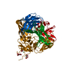
| Title | Crystal structure of Putative PhoU-like phosphate regulatory protein (BT4638) from BACTEROIDES THETAIOTAOMICRON VPI-5482 at 1.93 A resolution | ||||||
 Components Components | Putative PhoU-like phosphate regulatory protein | ||||||
 Keywords Keywords | phosphate-binding protein / BT4638 / Putative PhoU-like phosphate regulatory protein / Structural Genomics / Joint Center for Structural Genomics / JCSG / Protein Structure Initiative / PSI-2 | ||||||
| Function / homology |  Function and homology information Function and homology information: / Putative phosphate transport regulator / Protein of unknown function DUF47 / Phosphate transport system protein phou homolog 2; domain 2 / PhoU-like domain superfamily / Methane Monooxygenase Hydroxylase; Chain G, domain 1 / Up-down Bundle / Mainly Alpha Similarity search - Domain/homology | ||||||
| Biological species |  Bacteroides thetaiotaomicron (bacteria) Bacteroides thetaiotaomicron (bacteria) | ||||||
| Method |  X-RAY DIFFRACTION / SYNCHROTRON / MOLECULAR REPLACEMENT / molecular replacement / Resolution: 1.93 Å X-RAY DIFFRACTION / SYNCHROTRON / MOLECULAR REPLACEMENT / molecular replacement / Resolution: 1.93 Å | ||||||
 Authors Authors | Joint Center for Structural Genomics (JCSG) | ||||||
 Citation Citation | Journal: To be published Title: Crystal structure of Putative PhoU-like phosphate regulatory protein (BT4638) from BACTEROIDES THETAIOTAOMICRON VPI-5482 at 1.93 A resolution Authors: Joint Center for Structural Genomics (JCSG) | ||||||
| History |
|
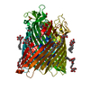
- Structure visualization
| Structure viewer | Molecule: MolmilJmol/JSmol |
|---|
- Downloads & links
-Download
| PDBx/mmCIF format | 3l39.cif.gz | 63 KB | Display | PDBx/mmCIF format |
|---|---|---|---|---|
| PDB format | pdb3l39.ent.gz | 45.7 KB | Display | PDB format |
| PDBx/mmJSON format | 3l39.json.gz | Tree view | PDBx/mmJSON format | |
| Others |  Other downloads Other downloads |
-Validation report
| Summary document | 3l39_validation.pdf.gz | 437.9 KB | Display | wwPDB validaton report |
|---|---|---|---|---|
| Full document | 3l39_full_validation.pdf.gz | 438.9 KB | Display | |
| Data in XML | 3l39_validation.xml.gz | 11.9 KB | Display | |
| Data in CIF | 3l39_validation.cif.gz | 17.3 KB | Display | |
| Arichive directory | https://data.pdbj.org/pub/pdb/validation_reports/l3/3l39ftp://data.pdbj.org/pub/pdb/validation_reports/l3/3l39 | HTTPS FTP |
-Related structure data
| Related structure data |  1xwmS  2iiuS S: Starting model for refinement |
|---|---|
| Similar structure data | |
| Other databases |


-Links
 PDBj
PDBj- Assembly
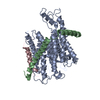
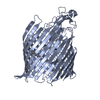
| Deposited unit | 
| ||||||||
|---|---|---|---|---|---|---|---|---|---|
| 1 | 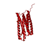
| ||||||||
| Unit cell |
| ||||||||
| Components on special symmetry positions |
|
-Components
| #1: Protein | Mass: 26499.213 Da / Num. of mol.: 1 Source method: isolated from a genetically manipulated source Source: (gene. exp.) Bacteroides thetaiotaomicron (bacteria)Strain: VPI-5482 / Gene: BT4638, BT_4638 / Plasmid: MH4a / Production host: | ||||
|---|---|---|---|---|---|
| #2: Chemical | ChemComp-PO4 /   Mass: 94.971 Da / Num. of mol.: 7 / Source method: obtained synthetically / Formula: PO4 Mass: 94.971 Da / Num. of mol.: 7 / Source method: obtained synthetically / Formula: PO4#3: Water | ChemComp-HOH / |  Mass: 18.015 Da / Num. of mol.: 187 / Source method: isolated from a natural source / Formula: H2O Mass: 18.015 Da / Num. of mol.: 187 / Source method: isolated from a natural source / Formula: H2OSequence details | THE CONSTRUCT WAS EXPRESSED WITH A PURIFICATI | |
-Experimental details
-Experiment
| Experiment | Method: X-RAY DIFFRACTION / Number of used crystals: 1 |
|---|
- Sample preparation
| Crystal | Density Matthews: 2.73 Å3/Da / Density % sol: 54.94 % |
|---|---|
| Crystal grow | Temperature: 277 K / Method: vapor diffusion, sitting drop / pH: 8.5 Details: 20.0% Glycerol, 1.6M NH4H2PO3, 0.1M TRIS pH 8.5, NANODROP, VAPOR DIFFUSION, SITTING DROP, temperature 277K |
-Data collection
| Diffraction | Mean temperature: 100 K |
|---|---|
| Diffraction source | Source: SYNCHROTRON / Site: SSRL  / Beamline: BL9-2 / Wavelength: 0.979245 / Beamline: BL9-2 / Wavelength: 0.979245 |
| Detector | Type: ADSC QUANTUM 315 / Detector: CCD / Date: Jul 18, 2004 / Details: Flat collimating mirror, toroid focusing mirror |
| Radiation | Monochromator: Double crystal monochromator / Protocol: SINGLE WAVELENGTH / Monochromatic (M) / Laue (L): M / Scattering type: x-ray |
| Radiation wavelength | Wavelength: 0.979245 Å / Relative weight: 1 |
| Reflection | Resolution: 1.93→44.972 Å / Num. obs: 18980 / % possible obs: 85.3 % / Redundancy: 5.3 % / Biso Wilson estimate: 16.512 Å2 / Rsym value: 0.071 |
-Phasing
| Phasing | Method: molecular replacement |
|---|
- Processing
| Software |
| |||||||||||||||||||||||||||||||||||||||||||||||||||||||||||||||||||||||||||||||||||||
|---|---|---|---|---|---|---|---|---|---|---|---|---|---|---|---|---|---|---|---|---|---|---|---|---|---|---|---|---|---|---|---|---|---|---|---|---|---|---|---|---|---|---|---|---|---|---|---|---|---|---|---|---|---|---|---|---|---|---|---|---|---|---|---|---|---|---|---|---|---|---|---|---|---|---|---|---|---|---|---|---|---|---|---|---|---|---|
| Refinement | Method to determine structure: MOLECULAR REPLACEMENT Starting model: PDB entry 2iiu,1xwm Resolution: 1.93→44.972 Å / Cor.coef. Fo:Fc: 0.949 / Cor.coef. Fo:Fc free: 0.934 / Occupancy max: 1 / Occupancy min: 0.33 / SU B: 2.679 / SU ML: 0.079 / Cross valid method: THROUGHOUT / σ(F): 0 / ESU R: 0.147 / ESU R Free: 0.133 / Stereochemistry target values: MAXIMUM LIKELIHOOD Details: 1. HYDROGENS HAVE BEEN ADDED IN THE RIDING POSITIONS. 2. PHOSPHATES (PO4) FROM THE CRYSTALLIZATION SOLUTION WERE MODELED INTO THE STRUCTURE.
| |||||||||||||||||||||||||||||||||||||||||||||||||||||||||||||||||||||||||||||||||||||
| Solvent computation | Ion probe radii: 0.8 Å / Shrinkage radii: 0.8 Å / VDW probe radii: 1.4 Å / Solvent model: MASK | |||||||||||||||||||||||||||||||||||||||||||||||||||||||||||||||||||||||||||||||||||||
| Displacement parameters | Biso max: 63.46 Å2 / Biso mean: 14.477 Å2 / Biso min: 3.05 Å2
| |||||||||||||||||||||||||||||||||||||||||||||||||||||||||||||||||||||||||||||||||||||
| Refinement step | Cycle: LAST / Resolution: 1.93→44.972 Å
| |||||||||||||||||||||||||||||||||||||||||||||||||||||||||||||||||||||||||||||||||||||
| Refine LS restraints |
| |||||||||||||||||||||||||||||||||||||||||||||||||||||||||||||||||||||||||||||||||||||
| LS refinement shell | Resolution: 1.93→1.98 Å / Total num. of bins used: 20
|
