 Movie
Movie Controller

[English] 日本語
 Yorodumi
- PDB-3kxg: Crystal structure of Z. mays CK2 kinase alpha subunit in complex ... -
+ Open data

- Basic information
| Entry | Database: PDB / ID: 3kxg | ||||||
|---|---|---|---|---|---|---|---|
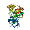
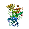
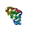
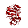
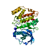
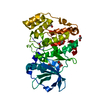
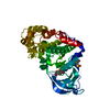
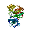
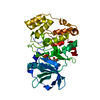
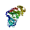
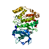
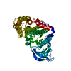
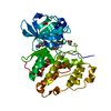
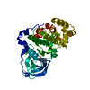
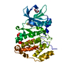
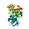
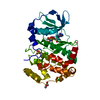
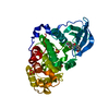
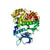
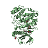
| Title | Crystal structure of Z. mays CK2 kinase alpha subunit in complex with the inhibitor 3,4,5,6,7-pentabromo-1H-indazole (K64) | ||||||
 Components Components | Casein kinase II subunit alpha | ||||||
 Keywords Keywords | Transferase/Transferase inhibitor / Protein kinase CK2-inhibitor complex / ATP-binding / Kinase / Nucleotide-binding / Serine/threonine-protein kinase / Transferase / Transferase-Transferase inhibitor complex | ||||||
| Function / homology |  Function and homology information Function and homology informationphotoperiodism / inflorescence development / protein kinase CK2 complex / non-specific serine/threonine protein kinase / regulation of cell cycle / protein serine kinase activity / protein serine/threonine kinase activity / DNA damage response / ATP binding / nucleus / cytosol Similarity search - Function | ||||||
| Biological species |  | ||||||
| Method |  X-RAY DIFFRACTION / SYNCHROTRON / Rigid body in an isomorphous cell / Resolution: 1.7 Å X-RAY DIFFRACTION / SYNCHROTRON / Rigid body in an isomorphous cell / Resolution: 1.7 Å | ||||||
 Authors Authors | Papinutto, E. / Franchin, C. / Battistutta, R. | ||||||
 Citation Citation | Journal: Curr Top Med Chem / Year: 2011 Title: ATP site-directed inhibitors of protein kinase CK2: an update. Authors: Sarno, S. / Papinutto, E. / Franchin, C. / Bain, J. / Elliott, M. / Meggio, F. / Kazimierczuk, Z. / Orzeszko, A. / Zanotti, G. / Battistutta, R. / Pinna, L.A. #1: Journal: Chem.Biol. / Year: 2005Title: Inspecting the structure-activity relationship of protein kinase CK2 inhibitors derived from tetrabromo-benzimidazole. Authors: Battistutta, R. / Mazzorana, M. / Sarno, S. / Kazimierczuk, Z. / Zanotti, G. / Pinna, L.A. #2: Journal: Chembiochem / Year: 2007Title: The ATP-binding site of protein kinase CK2 holds a positive electrostatic area and conserved water molecules. Authors: Battistutta, R. / Mazzorana, M. / Cendron, L. / Bortolato, A. / Sarno, S. / Kazimierczuk, Z. / Zanotti, G. / Moro, S. / Pinna, L.A. | ||||||
| History |
|
- Structure visualization
| Structure viewer | Molecule: MolmilJmol/JSmol |
|---|
- Downloads & links
-Download
| PDBx/mmCIF format | 3kxg.cif.gz | 88 KB | Display | PDBx/mmCIF format |
|---|---|---|---|---|
| PDB format | pdb3kxg.ent.gz | 64.9 KB | Display | PDB format |
| PDBx/mmJSON format | 3kxg.json.gz | Tree view | PDBx/mmJSON format | |
| Others |  Other downloads Other downloads |
-Validation report
| Arichive directory | https://data.pdbj.org/pub/pdb/validation_reports/kx/3kxgftp://data.pdbj.org/pub/pdb/validation_reports/kx/3kxg | HTTPS FTP |
|---|
-Related structure data


-Links
 PDBj
PDBj- Assembly
| Deposited unit | 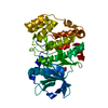
| ||||||||
|---|---|---|---|---|---|---|---|---|---|
| 1 |
| ||||||||
| Unit cell |
| ||||||||
| Components on special symmetry positions |
|
-Components
| #1: Protein | Mass: 38689.395 Da / Num. of mol.: 1 / Fragment: ALPHA SUBUNIT Source method: isolated from a genetically manipulated source Source: (gene. exp.)  References: UniProt: P28523, non-specific serine/threonine protein kinase |
|---|---|
| #2: Chemical | ChemComp-K6X /   Mass: 512.616 Da / Num. of mol.: 1 / Source method: obtained synthetically / Formula: C7HBr5N2 Mass: 512.616 Da / Num. of mol.: 1 / Source method: obtained synthetically / Formula: C7HBr5N2 |
| #3: Water | ChemComp-HOH /  Mass: 18.015 Da / Num. of mol.: 266 / Source method: isolated from a natural source / Formula: H2O Mass: 18.015 Da / Num. of mol.: 266 / Source method: isolated from a natural source / Formula: H2O |
| Has protein modification | Y |
-Experimental details
-Experiment
| Experiment | Method: X-RAY DIFFRACTION / Number of used crystals: 1 |
|---|
- Sample preparation
| Crystal | Density Matthews: 2.46 Å3/Da / Density % sol: 50.07 % |
|---|---|
| Crystal grow | Temperature: 293 K / Method: vapor diffusion, sitting drop / pH: 8 Details: 20% PEG 4000, 0.2M Na-acetate, 0.1M Tris, pH 8.0, VAPOR DIFFUSION, SITTING DROP, temperature 293K |
-Data collection
| Diffraction | Mean temperature: 100 K |
|---|---|
| Diffraction source | Source: SYNCHROTRON / Site: ESRF  / Beamline: ID29 / Wavelength: 0.972991 Å / Beamline: ID29 / Wavelength: 0.972991 Å |
| Detector | Type: ADSC QUANTUM 315r / Detector: CCD / Date: Mar 6, 2008 / Details: mirrors |
| Radiation | Monochromator: Si(111) / Protocol: SINGLE WAVELENGTH / Monochromatic (M) / Laue (L): M / Scattering type: x-ray |
| Radiation wavelength | Wavelength: 0.972991 Å / Relative weight: 1 |
| Reflection | Resolution: 1.7→69.34 Å / Num. all: 41479 / Num. obs: 41272 / % possible obs: 99.5 % / Redundancy: 4 % / Rmerge(I) obs: 0.059 / Rsym value: 0.059 / Net I/σ(I): 16.3 |
| Reflection shell | Resolution: 1.7→1.79 Å / Redundancy: 4 % / Rmerge(I) obs: 0.441 / Mean I/σ(I) obs: 2.3 / Rsym value: 0.441 / % possible all: 100 |
- Processing
| Software |
| ||||||||||||||||||||||||||||||||||||||||||||||||||||||||||||||||||||||||||||||||||||||||||||||||||||||||||||||||||||||||||||||||||||||||||||||||||||||||||||||||||||||||||
|---|---|---|---|---|---|---|---|---|---|---|---|---|---|---|---|---|---|---|---|---|---|---|---|---|---|---|---|---|---|---|---|---|---|---|---|---|---|---|---|---|---|---|---|---|---|---|---|---|---|---|---|---|---|---|---|---|---|---|---|---|---|---|---|---|---|---|---|---|---|---|---|---|---|---|---|---|---|---|---|---|---|---|---|---|---|---|---|---|---|---|---|---|---|---|---|---|---|---|---|---|---|---|---|---|---|---|---|---|---|---|---|---|---|---|---|---|---|---|---|---|---|---|---|---|---|---|---|---|---|---|---|---|---|---|---|---|---|---|---|---|---|---|---|---|---|---|---|---|---|---|---|---|---|---|---|---|---|---|---|---|---|---|---|---|---|---|---|---|---|---|---|
| Refinement | Method to determine structure: Rigid body in an isomorphous cell Resolution: 1.7→69.34 Å / Cor.coef. Fo:Fc: 0.956 / Cor.coef. Fo:Fc free: 0.928 / SU B: 6.099 / SU ML: 0.102 / Cross valid method: THROUGHOUT / ESU R: 0.124 / ESU R Free: 0.126 / Stereochemistry target values: MAXIMUM LIKELIHOOD / Details: HYDROGENS HAVE BEEN ADDED IN THE RIDING POSITIONS
| ||||||||||||||||||||||||||||||||||||||||||||||||||||||||||||||||||||||||||||||||||||||||||||||||||||||||||||||||||||||||||||||||||||||||||||||||||||||||||||||||||||||||||
| Solvent computation | Ion probe radii: 0.8 Å / Shrinkage radii: 0.8 Å / VDW probe radii: 1.2 Å / Solvent model: MASK | ||||||||||||||||||||||||||||||||||||||||||||||||||||||||||||||||||||||||||||||||||||||||||||||||||||||||||||||||||||||||||||||||||||||||||||||||||||||||||||||||||||||||||
| Displacement parameters | Biso mean: 19.519 Å2
| ||||||||||||||||||||||||||||||||||||||||||||||||||||||||||||||||||||||||||||||||||||||||||||||||||||||||||||||||||||||||||||||||||||||||||||||||||||||||||||||||||||||||||
| Refinement step | Cycle: LAST / Resolution: 1.7→69.34 Å
| ||||||||||||||||||||||||||||||||||||||||||||||||||||||||||||||||||||||||||||||||||||||||||||||||||||||||||||||||||||||||||||||||||||||||||||||||||||||||||||||||||||||||||
| Refine LS restraints |
| ||||||||||||||||||||||||||||||||||||||||||||||||||||||||||||||||||||||||||||||||||||||||||||||||||||||||||||||||||||||||||||||||||||||||||||||||||||||||||||||||||||||||||
| LS refinement shell | Resolution: 1.7→1.744 Å / Total num. of bins used: 20
|
