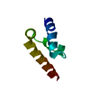
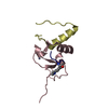
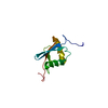
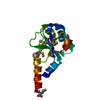
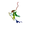
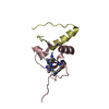
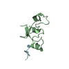
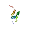
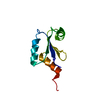
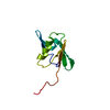
[English] 日本語
Yorodumi
- PDB-3kui: Crystal structure of C-terminal domain of PABPC1 in complex with ... -

+
Open data


ID or keywords:

Loading...

-
Basic information

Entry
Database: PDB / ID: 3kui
TitleCrystal structure of C-terminal domain of PABPC1 in complex with binding region of eRF3a
Components
  • GSPT1 protein
  • Polyadenylate-binding protein 1
KeywordsPROTEIN BINDING / protein-protein complex / Acetylation / Alternative splicing / Cytoplasm / Methylation / mRNA processing / mRNA splicing / Nucleus / Phosphoprotein / RNA-binding / Spliceosome / GTP-binding / Nucleotide-binding
Function / homology
Function and homology information


negative regulation of nuclear-transcribed mRNA catabolic process, nonsense-mediated decay / mCRD-mediated mRNA stability complex / translation release factor complex / translation activator activity / negative regulation of nuclear-transcribed mRNA catabolic process, deadenylation-dependent decay / CRD-mediated mRNA stabilization / regulation of translational termination / translation release factor activity / Z-decay: degradation of maternal mRNAs by zygotically expressed factors / Deadenylation of mRNA ...negative regulation of nuclear-transcribed mRNA catabolic process, nonsense-mediated decay / mCRD-mediated mRNA stability complex / translation release factor complex / translation activator activity / negative regulation of nuclear-transcribed mRNA catabolic process, deadenylation-dependent decay / CRD-mediated mRNA stabilization / regulation of translational termination / translation release factor activity / Z-decay: degradation of maternal mRNAs by zygotically expressed factors / Deadenylation of mRNA / protein methylation / positive regulation of cytoplasmic translation / mRNA stabilization / poly(A) binding / M-decay: degradation of maternal mRNAs by maternally stored factors / regulatory ncRNA-mediated gene silencing / poly(U) RNA binding / nuclear-transcribed mRNA catabolic process, nonsense-mediated decay / positive regulation of nuclear-transcribed mRNA catabolic process, deadenylation-dependent decay / positive regulation of nuclear-transcribed mRNA poly(A) tail shortening / Translation initiation complex formation / : / cell leading edge / Eukaryotic Translation Termination / Nonsense Mediated Decay (NMD) independent of the Exon Junction Complex (EJC) / L13a-mediated translational silencing of Ceruloplasmin expression / positive regulation of viral genome replication / Nonsense Mediated Decay (NMD) enhanced by the Exon Junction Complex (EJC) / translational termination / catalytic step 2 spliceosome / cytosolic ribosome / mRNA 3'-UTR binding / AUF1 (hnRNP D0) binds and destabilizes mRNA / Hydrolases; Acting on acid anhydrides; Acting on GTP to facilitate cellular and subcellular movement / G1/S transition of mitotic cell cycle / cytoplasmic ribonucleoprotein granule / mRNA splicing, via spliceosome / Regulation of expression of SLITs and ROBOs / cytoplasmic stress granule / lamellipodium / ribonucleoprotein complex / translation / focal adhesion / mRNA binding / GTPase activity / GTP binding / RNA binding / extracellular exosome / membrane / nucleus / cytosol / cytoplasm
Similarity search - Function
c-terminal domain of poly(a) binding protein / c-terminal domain of poly(a) binding protein / : / PABP, RNA recognition motif 2 / Polyadenylate binding protein, human types 1, 2, 3, 4 / Polyadenylate-binding protein/Hyperplastic disc protein / Poly-adenylate binding protein, unique domain / Poly(A)-binding protein C-terminal (PABC) domain profile. / C-terminal domain of Poly(A)-binding protein. Present also in Drosophila hyperplastics discs protein. / PABC (PABP) domain ...c-terminal domain of poly(a) binding protein / c-terminal domain of poly(a) binding protein / : / PABP, RNA recognition motif 2 / Polyadenylate binding protein, human types 1, 2, 3, 4 / Polyadenylate-binding protein/Hyperplastic disc protein / Poly-adenylate binding protein, unique domain / Poly(A)-binding protein C-terminal (PABC) domain profile. / C-terminal domain of Poly(A)-binding protein. Present also in Drosophila hyperplastics discs protein. / PABC (PABP) domain / RNA recognition motif domain, eukaryote / RNA recognition motif / Translation elongation factor EFTu/EF1A, C-terminal / Elongation factor Tu C-terminal domain / Translation elongation factor EF1A/initiation factor IF2gamma, C-terminal / Tr-type G domain, conserved site / Translational (tr)-type guanine nucleotide-binding (G) domain signature. / Translation elongation factor EFTu-like, domain 2 / Elongation factor Tu domain 2 / Translational (tr)-type GTP-binding domain / Elongation factor Tu GTP binding domain / Translational (tr)-type guanine nucleotide-binding (G) domain profile. / RNA recognition motif / RNA recognition motif / Eukaryotic RNA Recognition Motif (RRM) profile. / RNA recognition motif domain / RNA-binding domain superfamily / Translation protein, beta-barrel domain superfamily / Nucleotide-binding alpha-beta plait domain superfamily / P-loop containing nucleoside triphosphate hydrolase / Orthogonal Bundle / Mainly Alpha
Similarity search - Domain/homology
Polyadenylate-binding protein 1 / Eukaryotic peptide chain release factor GTP-binding subunit ERF3A / Eukaryotic peptide chain release factor GTP-binding subunit ERF3A
Similarity search - Component
Biological speciesHomo sapiens (human)
MethodX-RAY DIFFRACTION / MOLECULAR REPLACEMENT / Resolution: 2.3 Å
AuthorsKozlov, G. / Gehring, K.
CitationJournal: Plos One / Year: 2010
Title: Molecular basis of eRF3 recognition by the MLLE domain of poly(A)-binding protein.
Authors: Kozlov, G. / Gehring, K.
History
DepositionNov 27, 2009Deposition site: RCSB / Processing site: RCSB
Revision 1.0May 12, 2010Provider: repository / Type: Initial release
Revision 1.1Jul 13, 2011Group: Version format compliance
Revision 1.2Sep 6, 2023Group: Data collection / Database references ...Data collection / Database references / Derived calculations / Refinement description
Category: chem_comp_atom / chem_comp_bond ...chem_comp_atom / chem_comp_bond / database_2 / pdbx_initial_refinement_model / struct_ref_seq_dif / struct_site
Item: _database_2.pdbx_DOI / _database_2.pdbx_database_accession ..._database_2.pdbx_DOI / _database_2.pdbx_database_accession / _struct_ref_seq_dif.details / _struct_site.pdbx_auth_asym_id / _struct_site.pdbx_auth_comp_id / _struct_site.pdbx_auth_seq_id

-
Structure visualization

Structure viewerMolecule:
MolmilJmol/JSmol

Downloads & links

-
Assembly

Deposited unit
A: Polyadenylate-binding protein 1
B: GSPT1 protein
hetero molecules


Theoretical massNumber of molelcules
Total (without water)11,3194
Polymers11,1582
Non-polymers1612
Water28816
1


  • Idetical with deposited unit
  • defined by author&software
TypeNameSymmetry operationNumber
identity operation1_555x,y,z1
Buried area1550 Å2
ΔGint-47 kcal/mol
Surface area5770 Å2
MethodPISA
Unit cell
Length a, b, c (Å)37.432, 63.631, 32.226
Angle α, β, γ (deg.)90.00, 90.00, 90.00
Int Tables number18
Space group name H-MP21212
Components on special symmetry positions
IDModelComponents
11A-4-

HOH

-
Components

#1: Protein Polyadenylate-binding protein 1 / Poly(A)-binding protein 1 / PABP 1


Mass: 9421.909 Da / Num. of mol.: 1 / Fragment: C-terminal domain
Source method: isolated from a genetically manipulated source
Source: (gene. exp.) Homo sapiens (human) / Gene: PABPC1, PAB1, PABP1, PABPC2 / Plasmid: pGEX-6P-1 / Production host: Escherichia coli (E. coli) / Strain (production host): BL21 / References: UniProt: P11940
#2: Protein/peptide GSPT1 protein /


Mass: 1736.004 Da / Num. of mol.: 1 / Fragment: PABPC1-binding region / Source method: obtained synthetically / Details: chemically synthesized / Source: (synth.) Homo sapiens (human) / References: UniProt: Q96GF2, UniProt: P15170*PLUS
#3: Chemical ChemComp-ZN / ZINC ION


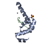
Mass: 65.409 Da / Num. of mol.: 1 / Source method: obtained synthetically / Formula: Zn
#4: Chemical ChemComp-SO4 / SULFATE ION / Sulfate


Mass: 96.063 Da / Num. of mol.: 1 / Source method: obtained synthetically / Formula: SO4
#5: Water ChemComp-HOH / water / Water


Mass: 18.015 Da / Num. of mol.: 16 / Source method: isolated from a natural source / Formula: H2O

-
Experimental details

-
Experiment

ExperimentMethod: X-RAY DIFFRACTION / Number of used crystals: 1

-
Sample preparation

Crystal growTemperature: 295 K / Method: vapor diffusion, hanging drop / pH: 5.4
Details: 2.1M ammonium sulfate, 0.2M sodium sulfate, 10mM zinc chloride, 0.1M sodium acetate, pH 5.4, VAPOR DIFFUSION, HANGING DROP, temperature 295K

-
Data collection

DiffractionMean temperature: 140 K
Diffraction sourceSource: ROTATING ANODE / Type: RIGAKU / Wavelength: 1.54178 Å
DetectorType: RIGAKU RAXIS IV++ / Detector: IMAGE PLATE / Date: Nov 5, 2009 / Details: mirrors
RadiationMonochromator: graphite / Protocol: SINGLE WAVELENGTH / Monochromatic (M) / Laue (L): M / Scattering type: x-ray
Radiation wavelengthWavelength: 1.54178 Å / Relative weight: 1
ReflectionResolution: 2.3→50 Å / Num. all: 3570 / Num. obs: 3559 / % possible obs: 99.7 % / Observed criterion σ(F): 1 / Observed criterion σ(I): 1 / Redundancy: 7 % / Rmerge(I) obs: 0.094 / Net I/σ(I): 19.3
Reflection shellResolution: 2.3→2.34 Å / Redundancy: 5.8 % / Rmerge(I) obs: 0.415 / Mean I/σ(I) obs: 3.8 / Num. unique all: 235 / % possible all: 98.8

-
Processing

Software
NameVersionClassification
CrystalCleardata collection
PHASERphasing
CNS1.1refinement
HKL-2000data reduction
HKL-2000data scaling
RefinementMethod to determine structure: MOLECULAR REPLACEMENT
Starting model: pdb entry 1I2T
Resolution: 2.3→50 Å / Cross valid method: THROUGHOUT / σ(F): 1 / Stereochemistry target values: MAXIMUM LIKELIHOOD
RfactorNum. reflection% reflectionSelection details
Rfree0.255 191 -RANDOM
Rwork0.247 ---
obs0.248 3559 98.8 %-
all-3570 --
Displacement parametersBiso mean: 46.44 Å2
Refinement stepCycle: LAST / Resolution: 2.3→50 Å
ProteinNucleic acidLigandSolventTotal
Num. atoms707 0 6 16 729
Refine LS restraints
Refine-IDTypeDev ideal
X-RAY DIFFRACTIONc_bond_d0.007148
X-RAY DIFFRACTIONc_angle_deg1.15299
LS refinement shellResolution: 2.3→2.34 Å /
Num. reflection% reflection
Rfree17 -
obs424 98.8 %

+
About Yorodumi

-
News

-
Feb 9, 2022. New format data for meta-information of EMDB entries

New format data for meta-information of EMDB entries

  • Version 3 of the EMDB header file is now the official format.
  • The previous official version 1.9 will be removed from the archive.

Related info.:EMDB header

External links:wwPDB to switch to version 3 of the EMDB data model

-
Aug 12, 2020. Covid-19 info

Covid-19 info

URL: https://pdbj.org/emnavi/covid19.php

New page: Covid-19 featured information page in EM Navigator.

Related info.:Covid-19 info / Mar 5, 2020. Novel coronavirus structure data

+
Mar 5, 2020. Novel coronavirus structure data

Novel coronavirus structure data

Related info.:Yorodumi Speices / Aug 12, 2020. Covid-19 info

External links:COVID-19 featured content - PDBj / Molecule of the Month (242):Coronavirus Proteases

+
Jan 31, 2019. EMDB accession codes are about to change! (news from PDBe EMDB page)

EMDB accession codes are about to change! (news from PDBe EMDB page)

  • The allocation of 4 digits for EMDB accession codes will soon come to an end. Whilst these codes will remain in use, new EMDB accession codes will include an additional digit and will expand incrementally as the available range of codes is exhausted. The current 4-digit format prefixed with “EMD-” (i.e. EMD-XXXX) will advance to a 5-digit format (i.e. EMD-XXXXX), and so on. It is currently estimated that the 4-digit codes will be depleted around Spring 2019, at which point the 5-digit format will come into force.
  • The EM Navigator/Yorodumi systems omit the EMD- prefix.

Related info.:Q: What is EMD? / ID/Accession-code notation in Yorodumi/EM Navigator

External links:EMDB Accession Codes are Changing Soon! / Contact to PDBj

+
Jul 12, 2017. Major update of PDB

Major update of PDB

  • wwPDB released updated PDB data conforming to the new PDBx/mmCIF dictionary.
  • This is a major update changing the version number from 4 to 5, and with Remediation, in which all the entries are updated.
  • In this update, many items about electron microscopy experimental information are reorganized (e.g. em_software).
  • Now, EM Navigator and Yorodumi are based on the updated data.

External links:wwPDB Remediation / Enriched Model Files Conforming to OneDep Data Standards Now Available in the PDB FTP Archive

-
Yorodumi

Thousand views of thousand structures

  • Yorodumi is a browser for structure data from EMDB, PDB, SASBDB, etc.
  • This page is also the successor to EM Navigator detail page, and also detail information page/front-end page for Omokage search.
  • The word "yorodu" (or yorozu) is an old Japanese word meaning "ten thousand". "mi" (miru) is to see.

Related info.:EMDB / PDB / SASBDB / Comparison of 3 databanks / Yorodumi Search / Aug 31, 2016. New EM Navigator & Yorodumi / Yorodumi Papers / Jmol/JSmol / Function and homology information / Changes in new EM Navigator and Yorodumi

Read more