 Movie
Movie Controller

[English] 日本語
 Yorodumi
- PDB-3ksi: structure of fRMsr of Staphylococcus aureus (complex with 2-propanol) -
+ Open data

- Basic information
| Entry | Database: PDB / ID: 3ksi | ||||||
|---|---|---|---|---|---|---|---|
| Title | structure of fRMsr of Staphylococcus aureus (complex with 2-propanol) | ||||||
 Components Components | Putative uncharacterized protein | ||||||
 Keywords Keywords | OXIDOREDUCTASE / fRMsr / free-Met-R-SO | ||||||
| Function / homology |  Function and homology information Function and homology information | ||||||
| Biological species |   Staphylococcus aureus (bacteria) Staphylococcus aureus (bacteria) | ||||||
| Method |  X-RAY DIFFRACTION / SYNCHROTRON / MOLECULAR REPLACEMENT / Resolution: 1.7 Å X-RAY DIFFRACTION / SYNCHROTRON / MOLECULAR REPLACEMENT / Resolution: 1.7 Å | ||||||
 Authors Authors | Bong, S.M. / Chi, Y.M. | ||||||
 Citation Citation | Journal: To be Published Title: structure of fRMsr of Staphylococcus aureus Authors: Bong, S.M. / Chi, Y.M. | ||||||
| History |
|
- Structure visualization
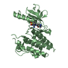
| Structure viewer | Molecule: MolmilJmol/JSmol |
|---|
- Downloads & links
-Download
| PDBx/mmCIF format | 3ksi.cif.gz | 45.6 KB | Display | PDBx/mmCIF format |
|---|---|---|---|---|
| PDB format | pdb3ksi.ent.gz | 31 KB | Display | PDB format |
| PDBx/mmJSON format | 3ksi.json.gz | Tree view | PDBx/mmJSON format | |
| Others |  Other downloads Other downloads |
-Validation report
| Summary document | 3ksi_validation.pdf.gz | 448.8 KB | Display | wwPDB validaton report |
|---|---|---|---|---|
| Full document | 3ksi_full_validation.pdf.gz | 450.8 KB | Display | |
| Data in XML | 3ksi_validation.xml.gz | 9.3 KB | Display | |
| Data in CIF | 3ksi_validation.cif.gz | 12.2 KB | Display | |
| Arichive directory | https://data.pdbj.org/pub/pdb/validation_reports/ks/3ksiftp://data.pdbj.org/pub/pdb/validation_reports/ks/3ksi | HTTPS FTP |
-Related structure data
| Related structure data |  3ksfC  1vhmS C: citing same article ( S: Starting model for refinement |
|---|---|
| Similar structure data |


-Links
 PDBj
PDBj


- Assembly
| Deposited unit | 
| ||||||||
|---|---|---|---|---|---|---|---|---|---|
| 1 | 
| ||||||||
| Unit cell |
|
-Components
| #1: Protein | Mass: 17944.475 Da / Num. of mol.: 1 Source method: isolated from a genetically manipulated source Source: (gene. exp.) Staphylococcus aureus (bacteria) / Strain: MRSA252 / Gene: SAR1796 / Plasmid: pET28a / Production host: References: UniProt: Q6GFY9, UniProt: A0A7U7EVB7*PLUS, L-methionine (R)-S-oxide reductase |
|---|---|
| #2: Chemical | ChemComp-IPA /   Mass: 60.095 Da / Num. of mol.: 1 / Source method: obtained synthetically / Formula: C3H8O / Comment: alkaloid*YM Mass: 60.095 Da / Num. of mol.: 1 / Source method: obtained synthetically / Formula: C3H8O / Comment: alkaloid*YM |
| #3: Chemical | ChemComp-SO4 /   Mass: 96.063 Da / Num. of mol.: 1 / Source method: obtained synthetically / Formula: SO4 Mass: 96.063 Da / Num. of mol.: 1 / Source method: obtained synthetically / Formula: SO4 |
| #4: Water | ChemComp-HOH /  Mass: 18.015 Da / Num. of mol.: 109 / Source method: isolated from a natural source / Formula: H2O Mass: 18.015 Da / Num. of mol.: 109 / Source method: isolated from a natural source / Formula: H2O |
-Experimental details
-Experiment
| Experiment | Method: X-RAY DIFFRACTION / Number of used crystals: 1 |
|---|
- Sample preparation
| Crystal | Density Matthews: 2.88 Å3/Da / Density % sol: 57.3 % |
|---|---|
| Crystal grow | Temperature: 295 K / Method: vapor diffusion, hanging drop Details: 2M ammonium sulfate, 10% isopropanol, VAPOR DIFFUSION, HANGING DROP, temperature 295K |
-Data collection
| Diffraction | Mean temperature: 100 K |
|---|---|
| Diffraction source | Source: SYNCHROTRON / Site: PAL/PLS  / Beamline: 4A / Wavelength: 1.2386 Å / Beamline: 4A / Wavelength: 1.2386 Å |
| Detector | Type: ADSC QUANTUM 210 / Detector: CCD / Date: Mar 31, 2008 |
| Radiation | Monochromator: diamond / Protocol: SINGLE WAVELENGTH / Monochromatic (M) / Laue (L): M / Scattering type: x-ray |
| Radiation wavelength | Wavelength: 1.2386 Å / Relative weight: 1 |
| Reflection | Resolution: 1.7→50 Å / Num. obs: 23645 / % possible obs: 94.8 % / Observed criterion σ(F): 0 / Observed criterion σ(I): 0 |
- Processing
| Software |
| ||||||||||||||||||||
|---|---|---|---|---|---|---|---|---|---|---|---|---|---|---|---|---|---|---|---|---|---|
| Refinement | Method to determine structure: MOLECULAR REPLACEMENT Starting model: PDB ENTRY 1VHM Resolution: 1.7→44.92 Å / σ(F): 0 / Stereochemistry target values: Engh & Huber
| ||||||||||||||||||||
| Solvent computation | Bsol: 53.444 Å2 | ||||||||||||||||||||
| Displacement parameters | Biso mean: 23.891 Å2
| ||||||||||||||||||||
| Refine analyze |
| ||||||||||||||||||||
| Refinement step | Cycle: LAST / Resolution: 1.7→44.92 Å
| ||||||||||||||||||||
| Refine LS restraints |
| ||||||||||||||||||||
| LS refinement shell | Resolution: 1.7→1.81 Å / Rfactor Rfree error: 0.014
|
