 Movie
Movie Controller

[English] 日本語
 Yorodumi
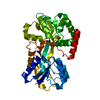
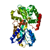
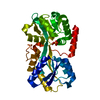
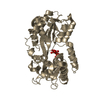
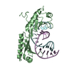
- PDB-3kn8: Crystal Structure of Haemophilus influenzae Y196A mutant Holo Fer... -
+ Open data

- Basic information
| Entry | Database: PDB / ID: 3kn8 | ||||||
|---|---|---|---|---|---|---|---|
| Title | Crystal Structure of Haemophilus influenzae Y196A mutant Holo Ferric ion-Binding Protein A | ||||||
 Components Components | Iron-utilization periplasmic protein | ||||||
 Keywords Keywords | METAL BINDING PROTEIN / iron binding protein / Iron / Iron transport / Metal-binding / Transport | ||||||
| Function / homology |  Function and homology information Function and homology informationiron ion transport / transmembrane transport / periplasmic space / metal ion binding Similarity search - Function | ||||||
| Biological species |  Haemophilus influenzae (bacteria) Haemophilus influenzae (bacteria) | ||||||
| Method |  X-RAY DIFFRACTION / MOLECULAR REPLACEMENT / molecular replacement / Resolution: 1.89 Å X-RAY DIFFRACTION / MOLECULAR REPLACEMENT / molecular replacement / Resolution: 1.89 Å | ||||||
 Authors Authors | Shouldice, S.R. / Schryvers, A.B. | ||||||
 Citation Citation | Journal: Biochem.J. / Year: 2010 Title: The role of vicinal tyrosine residues in the function of Haemophilus influenzae ferric binding protein A. Authors: Khambati, H.K. / Moraes, T.F. / Singh, J. / Shouldice, S.R. / Yu, R.H. / Schryvers, A.B. | ||||||
| History |
|
- Structure visualization
| Structure viewer | Molecule: MolmilJmol/JSmol |
|---|
- Downloads & links
-Download
| PDBx/mmCIF format | 3kn8.cif.gz | 76.9 KB | Display | PDBx/mmCIF format |
|---|---|---|---|---|
| PDB format | pdb3kn8.ent.gz | 55.2 KB | Display | PDB format |
| PDBx/mmJSON format | 3kn8.json.gz | Tree view | PDBx/mmJSON format | |
| Others |  Other downloads Other downloads |
-Validation report
| Arichive directory | https://data.pdbj.org/pub/pdb/validation_reports/kn/3kn8ftp://data.pdbj.org/pub/pdb/validation_reports/kn/3kn8 | HTTPS FTP |
|---|
-Related structure data
| Related structure data |  3kn7C  3od7C  3odbC 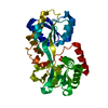 1d9vS C: citing same article ( S: Starting model for refinement |
|---|---|
| Similar structure data |


-Links
 PDBj
PDBj


- Assembly
| Deposited unit | 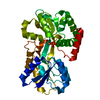
| |||||||||
|---|---|---|---|---|---|---|---|---|---|---|
| 1 |
| |||||||||
| Unit cell |
| |||||||||
| Components on special symmetry positions |
|
-Components
| #1: Protein | Mass: 33677.156 Da / Num. of mol.: 1 / Mutation: Y196A Source method: isolated from a genetically manipulated source Source: (gene. exp.) Haemophilus influenzae (bacteria) / Gene: fbp, fbpA, HI0097, hitA / Plasmid: pT7-7 / Production host: |
|---|---|
| #2: Chemical | ChemComp-FE /   Mass: 55.845 Da / Num. of mol.: 1 / Source method: obtained synthetically / Formula: Fe Mass: 55.845 Da / Num. of mol.: 1 / Source method: obtained synthetically / Formula: Fe |
| #3: Chemical | ChemComp-PO4 /   Mass: 94.971 Da / Num. of mol.: 1 / Source method: obtained synthetically / Formula: PO4 Mass: 94.971 Da / Num. of mol.: 1 / Source method: obtained synthetically / Formula: PO4 |
| #4: Water | ChemComp-HOH /  Mass: 18.015 Da / Num. of mol.: 245 / Source method: isolated from a natural source / Formula: H2O Mass: 18.015 Da / Num. of mol.: 245 / Source method: isolated from a natural source / Formula: H2O |
-Experimental details
-Experiment
| Experiment | Method: X-RAY DIFFRACTION / Number of used crystals: 1 |
|---|
- Sample preparation
| Crystal | Density Matthews: 1.97 Å3/Da / Density % sol: 37.53 % |
|---|---|
| Crystal grow | Temperature: 293 K / Method: vapor diffusion, hanging drop / pH: 8.5 Details: 28% PEG 550 MME, 0.1M Tris pH 8.5, VAPOR DIFFUSION, HANGING DROP, temperature 293K |
-Data collection
| Diffraction | Mean temperature: 100 K |
|---|---|
| Diffraction source | Source: ROTATING ANODE / Type: RIGAKU / Wavelength: 1.5418 Å |
| Detector | Type: BRUKER SMART 6000 / Detector: CCD / Date: Jul 6, 2002 |
| Radiation | Monochromator: Yale mirrors / Protocol: SINGLE WAVELENGTH / Monochromatic (M) / Laue (L): M / Scattering type: x-ray |
| Radiation wavelength | Wavelength: 1.5418 Å / Relative weight: 1 |
| Reflection | Resolution: 1.89→62 Å / Num. all: 21800 / Num. obs: 20126 / % possible obs: 92.32 % / Observed criterion σ(F): 0 / Observed criterion σ(I): 0 / Redundancy: 3.05 % / Biso Wilson estimate: 20.802 Å2 / Rsym value: 0.049 / Net I/σ(I): 9.94 |
| Reflection shell | Resolution: 1.9→2.03 Å / Mean I/σ(I) obs: 2.02 / Rsym value: 0.2838 / % possible all: 92.32 |
-Phasing
| Phasing | Method: molecular replacement |
|---|
- Processing
| Software |
| ||||||||||||||||||||||||||||||||||||||||||||||||||||||||||||||||||||||||||||||||||||||||||||||||||||
|---|---|---|---|---|---|---|---|---|---|---|---|---|---|---|---|---|---|---|---|---|---|---|---|---|---|---|---|---|---|---|---|---|---|---|---|---|---|---|---|---|---|---|---|---|---|---|---|---|---|---|---|---|---|---|---|---|---|---|---|---|---|---|---|---|---|---|---|---|---|---|---|---|---|---|---|---|---|---|---|---|---|---|---|---|---|---|---|---|---|---|---|---|---|---|---|---|---|---|---|---|---|
| Refinement | Method to determine structure: MOLECULAR REPLACEMENT Starting model: PDB ENTRY 1D9V Resolution: 1.89→62 Å / Cor.coef. Fo:Fc: 0.949 / Cor.coef. Fo:Fc free: 0.916 / Occupancy max: 1 / Occupancy min: 0.5 / SU B: 4.044 / SU ML: 0.116 / Cross valid method: THROUGHOUT / σ(F): 0 / ESU R: 0.194 / ESU R Free: 0.17 / Stereochemistry target values: MAXIMUM LIKELIHOOD / Details: HYDROGENS HAVE BEEN ADDED IN THE RIDING POSITIONS
| ||||||||||||||||||||||||||||||||||||||||||||||||||||||||||||||||||||||||||||||||||||||||||||||||||||
| Solvent computation | Ion probe radii: 0.8 Å / Shrinkage radii: 0.8 Å / VDW probe radii: 1.4 Å / Solvent model: BABINET MODEL WITH MASK | ||||||||||||||||||||||||||||||||||||||||||||||||||||||||||||||||||||||||||||||||||||||||||||||||||||
| Displacement parameters | Biso max: 60.81 Å2 / Biso mean: 20.673 Å2 / Biso min: 5.96 Å2
| ||||||||||||||||||||||||||||||||||||||||||||||||||||||||||||||||||||||||||||||||||||||||||||||||||||
| Refinement step | Cycle: LAST / Resolution: 1.89→62 Å
| ||||||||||||||||||||||||||||||||||||||||||||||||||||||||||||||||||||||||||||||||||||||||||||||||||||
| Refine LS restraints |
| ||||||||||||||||||||||||||||||||||||||||||||||||||||||||||||||||||||||||||||||||||||||||||||||||||||
| LS refinement shell | Resolution: 1.894→1.943 Å / Total num. of bins used: 20
|
