Resolution: 2.8→28.56 Å / Rfactor Rfree error: 0.012 / Data cutoff high absF: 1302833.1 / Data cutoff low absF: 0 / Isotropic thermal model: RESTRAINED / Cross valid method: THROUGHOUT / σ(F): 0 / Stereochemistry target values: Engh & Huber Details: RESOLUTION-DEPENDENT WEIGHTING SCHEME OTHER REFINEMENT REMARKS: BULK SOLVENT MODEL USED RESIDUES AT THE N-TERMINUS (MET 222 THROUGH GLN 230), C-TERMINUS (GLU 502 THROUGH PRO 509) AND TWO IN ...Details: RESOLUTION-DEPENDENT WEIGHTING SCHEME OTHER REFINEMENT REMARKS: BULK SOLVENT MODEL USED RESIDUES AT THE N-TERMINUS (MET 222 THROUGH GLN 230), C-TERMINUS (GLU 502 THROUGH PRO 509) AND TWO IN THE ACTIVATION LOOP (GLY 399 AND ALA 400) HAVE WEAK OR NON-EXISTENT ELECTRON DENSITY. THE RESIDUES AT THE TERMINI ARE NOT INCLUDED IN REFINEMENT. THE FOLLOWING RESIDUES HAVE SIDE CHAIN ATOMS WHICH ARE POORLY DEFINED BY ELECTRON DENSITY BUT THEY HAVE BEEN INCLUDED IN REFINEMENT: GLU 237 (ENTIRE SIDE CHAIN), LYS 246 (BEYOND CG), LYS 276 (BEYOND CD), GLN 309 (BEYOND CB), GLU 390 (ENTIRE SIDE CHAIN), ASN 392 (BEYOND CB), ARG 397 (ENTIRE SIDE CHAIN), GLU 398 (BEYOND CB), LYS 401 (BEYOND CB), ARG 438 (BEYOND CD) AND ASN 464 (ENTIRE SIDE CHAIN).
In the structure databanks used in Yorodumi, some data are registered as the other names, "COVID-19 virus" and "2019-nCoV". Here are the details of the virus and the list of structure data.
Jan 31, 2019. EMDB accession codes are about to change! (news from PDBe EMDB page)
EMDB accession codes are about to change! (news from PDBe EMDB page)
The allocation of 4 digits for EMDB accession codes will soon come to an end. Whilst these codes will remain in use, new EMDB accession codes will include an additional digit and will expand incrementally as the available range of codes is exhausted. The current 4-digit format prefixed with “EMD-” (i.e. EMD-XXXX) will advance to a 5-digit format (i.e. EMD-XXXXX), and so on. It is currently estimated that the 4-digit codes will be depleted around Spring 2019, at which point the 5-digit format will come into force.
The EM Navigator/Yorodumi systems omit the EMD- prefix.
Related info.:Q: What is EMD? / ID/Accession-code notation in Yorodumi/EM Navigator
Yorodumi is a browser for structure data from EMDB, PDB, SASBDB, etc.
This page is also the successor to EM Navigator detail page, and also detail information page/front-end page for Omokage search.
The word "yorodu" (or yorozu) is an old Japanese word meaning "ten thousand". "mi" (miru) is to see.
Related info.:EMDB / PDB / SASBDB / Comparison of 3 databanks / Yorodumi Search / Aug 31, 2016. New EM Navigator & Yorodumi / Yorodumi Papers / Jmol/JSmol / Function and homology information / Changes in new EM Navigator and Yorodumi
 Movie
Movie Controller


 Open data
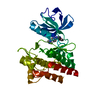
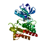
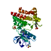
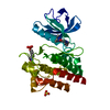
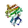
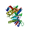
 Basic information
 Components
 Keywords
 Function and homology information
 Homo sapiens (human)
 X-RAY DIFFRACTION /
  Authors
 Citation
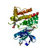
 Structure visualization
 Downloads & links
 Other downloads

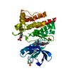
 PDBj
PDBj


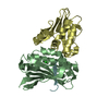
 Assembly
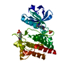

 Spodoptera frugiperda (fall armyworm) / Strain (production host): SF9
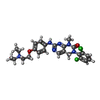
 Mass: 515.435 Da / Num. of mol.: 1 / Source method: obtained synthetically / Formula: C25H28Cl2N6O2
 Mass: 96.063 Da / Num. of mol.: 1 / Source method: obtained synthetically / Formula: SO4
 Mass: 18.015 Da / Num. of mol.: 115 / Source method: isolated from a natural source / Formula: H2O
 Sample preparation
 Processing