Monochromator: Double crystal monochromator / Protocol: SINGLE WAVELENGTH / Monochromatic (M) / Laue (L): M / Scattering type: x-ray
Radiation wavelength
Wavelength: 0.9792 Å / Relative weight: 1
Reflection
Resolution: 2.3→29.54 Å / Num. obs: 14896 / % possible obs: 99.6 % / Redundancy: 7.4 % / Biso Wilson estimate: 32.531 Å2 / Rmerge(I) obs: 0.22 / Rsym value: 0.22 / Net I/σ(I): 10.2
Reflection shell
Rmerge(I) obs: 0.013 / Diffraction-ID: 1
Resolution (Å)
Redundancy (%)
Mean I/σ(I) obs
Num. measured all
Num. unique all
Rsym value
% possible all
2.3-2.36
7.4
1.8
8211
1103
1.252
99
2.36-2.42
7.5
2.2
7917
1061
1.057
99.7
2.42-2.49
7.5
2.3
7638
1022
0.968
99.4
2.49-2.57
7.4
2.7
7293
979
0.825
99.4
2.57-2.66
7.4
3.3
7281
984
0.684
99.4
2.66-2.75
7.4
3.8
6834
924
0.588
99.5
2.75-2.85
7.5
4.2
6886
922
0.532
99.9
2.85-2.97
7.4
5.4
6442
868
0.402
99.6
2.97-3.1
7.5
7.1
6288
843
0.327
99.5
3.1-3.25
7.4
8.8
5881
796
0.257
99.7
3.25-3.43
7.4
11.6
5776
783
0.191
99.8
3.43-3.64
7.4
15.5
5313
722
0.142
99.8
3.64-3.89
7.3
18.4
5014
684
0.118
99.8
3.89-4.2
7.3
21.4
4775
657
0.099
99.8
4.2-4.6
7.3
25.8
4239
583
0.082
99.8
4.6-5.14
7.2
27.3
3994
551
0.074
99.9
5.14-5.94
7.1
22.2
3459
485
0.095
99.9
5.94-7.27
7.1
21.8
2902
410
0.103
99.9
7.27-10.29
6.8
31
2275
334
0.069
99.9
10.29-29.54
6.3
37.5
1160
185
0.056
95.8
-
Phasing
Phasing
Method: SAD
-
Processing
Software
Name
Version
Classification
NB
REFMAC
5.5.0053
refinement
PHENIX
refinement
SHELX
phasing
MolProbity
3beta29
modelbuilding
SCALA
3.2.5
datascaling
PDB_EXTRACT
3.006
dataextraction
MOSFLM
datareduction
SHELXD
phasing
autoSHARP
phasing
Refinement
Method to determine structure: SAD / Resolution: 2.3→29.54 Å / Cor.coef. Fo:Fc: 0.961 / Cor.coef. Fo:Fc free: 0.946 / Occupancy max: 1 / Occupancy min: 0.5 / SU B: 11.04 / SU ML: 0.122 / TLS residual ADP flag: LIKELY RESIDUAL / Cross valid method: THROUGHOUT / σ(F): 0 / ESU R: 0.212 / ESU R Free: 0.177 Stereochemistry target values: MAXIMUM LIKELIHOOD WITH PHASES Details: 1. HYDROGENS HAVE BEEN ADDED IN THE RIDING POSITIONS. 2. ATOM RECORD CONTAINS RESIDUAL B FACTORS ONLY. 3. A MET-INHIBITION PROTOCOL WAS USED FOR SELENOMETHIONINE INCORPORATION DURING PROTEIN ...Details: 1. HYDROGENS HAVE BEEN ADDED IN THE RIDING POSITIONS. 2. ATOM RECORD CONTAINS RESIDUAL B FACTORS ONLY. 3. A MET-INHIBITION PROTOCOL WAS USED FOR SELENOMETHIONINE INCORPORATION DURING PROTEIN EXPRESSION. THE OCCUPANCY OF THE SE ATOMS IN THE MSE RESIDUES WAS REDUCED TO 0.75 TO ACCOUNT FOR THE REDUCED SCATTERING POWER DUE TO PARTIAL S-MET INCORPORATION. 4. TWO ZINC IONS HAVE BEEN MODELED AT THE PUTATIVE ACTIVE SITE BASED ON AN X-RAY FLUORESCENCE EXCITATION SCAN SPECTRUM AND ANOMALOUS DIFFERENCE FOURIER PEAKS. 5. AN UNIDENTIFIED LIGAND (UNL) HAS BEEN MODELED AT THE PUTATIVE ACTIVE SITE COORDINATING THE ZINC SITES. 6. ACETATE (ACT) AND GLYCEROL (GOL) FROM THE CRYSTALLIZATION/ CRYOPROTECTANT SOLUTIONS HAVE BEEN MODELED INTO THE SOLVENT STRUCTURE. 7. TLS GROUPS WERE ASSIGNED WITH THE AID OF THE TLSMD SERVER.
Rfactor
Num. reflection
% reflection
Selection details
Rfree
0.204
750
5 %
RANDOM
Rwork
0.164
-
-
-
obs
0.166
14894
99.41 %
-
Solvent computation
Ion probe radii: 0.8 Å / Shrinkage radii: 0.8 Å / VDW probe radii: 1.4 Å / Solvent model: MASK
In the structure databanks used in Yorodumi, some data are registered as the other names, "COVID-19 virus" and "2019-nCoV". Here are the details of the virus and the list of structure data.
Jan 31, 2019. EMDB accession codes are about to change! (news from PDBe EMDB page)
EMDB accession codes are about to change! (news from PDBe EMDB page)
The allocation of 4 digits for EMDB accession codes will soon come to an end. Whilst these codes will remain in use, new EMDB accession codes will include an additional digit and will expand incrementally as the available range of codes is exhausted. The current 4-digit format prefixed with “EMD-” (i.e. EMD-XXXX) will advance to a 5-digit format (i.e. EMD-XXXXX), and so on. It is currently estimated that the 4-digit codes will be depleted around Spring 2019, at which point the 5-digit format will come into force.
The EM Navigator/Yorodumi systems omit the EMD- prefix.
Related info.:Q: What is EMD? / ID/Accession-code notation in Yorodumi/EM Navigator
Yorodumi is a browser for structure data from EMDB, PDB, SASBDB, etc.
This page is also the successor to EM Navigator detail page, and also detail information page/front-end page for Omokage search.
The word "yorodu" (or yorozu) is an old Japanese word meaning "ten thousand". "mi" (miru) is to see.
Related info.:EMDB / PDB / SASBDB / Comparison of 3 databanks / Yorodumi Search / Aug 31, 2016. New EM Navigator & Yorodumi / Yorodumi Papers / Jmol/JSmol / Function and homology information / Changes in new EM Navigator and Yorodumi
 Movie
Movie Controller

 Yorodumi
 Open data
 Basic information
 Components
 Keywords
 Function and homology information
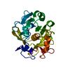
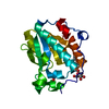
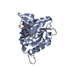
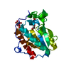
 Parabacteroides distasonis ATCC 8503 (bacteria)
 X-RAY DIFFRACTION /
  Authors
 Citation
 Structure visualization
 Downloads & links
 Other downloads


 PDBj
PDBj

 Assembly


 Mass: 65.409 Da / Num. of mol.: 2 / Source method: obtained synthetically / Formula: Zn
 Mass: 59.044 Da / Num. of mol.: 1 / Source method: obtained synthetically / Formula: C2H3O2
 Mass: 92.094 Da / Num. of mol.: 4 / Source method: obtained synthetically / Formula: C3H8O3
 Sample preparation
 / Beamline: BL9-2 / Wavelength: 0.9792
  Processing