 Movie
Movie Controller

[English] 日本語
 Yorodumi
- PDB-3khm: Crystal structure of sterol 14alpha-demethylase (CYP51) from Tryp... -
+ Open data

- Basic information
| Entry | Database: PDB / ID: 3khm | ||||||
|---|---|---|---|---|---|---|---|
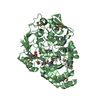
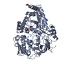
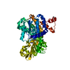
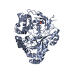
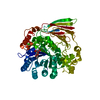
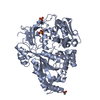
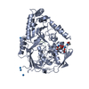
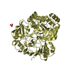
| Title | Crystal structure of sterol 14alpha-demethylase (CYP51) from Trypanosoma cruzi in complex with inhibitor fluconazole | ||||||
 Components Components | Sterol 14 alpha-demethylase | ||||||
 Keywords Keywords | OXIDOREDUCTASE / STEROL 14-ALPHA DEMETHYLASE / CYP51 / CYTOCHROME P450 / HEME / MONOOXYGENASE / ENDOPLASMIC RETICULUM / TRANSMEMBRANE PROTEIN / STEROL BIOSYNTHESIS / LIPIDS / MEMBRANE / IRON / HEME THIOLATE PROTEIN | ||||||
| Function / homology |  Function and homology information Function and homology informationsterol 14-demethylase activity / sterol biosynthetic process / sterol 14alpha-demethylase / iron ion binding / heme binding / membrane Similarity search - Function | ||||||
| Biological species |  | ||||||
| Method |  X-RAY DIFFRACTION / SYNCHROTRON / MOLECULAR REPLACEMENT / Resolution: 2.85 Å X-RAY DIFFRACTION / SYNCHROTRON / MOLECULAR REPLACEMENT / Resolution: 2.85 Å | ||||||
 Authors Authors | Lepesheva, G.I. / Hargrove, T.Y. / Anderson, S. / Wawrzak, Z. / Waterman, M.R. | ||||||
 Citation Citation | Journal: J.Biol.Chem. / Year: 2010 Title: Structural Insights into Inhibition of Sterol 14{alpha}-Demethylase in the Human Pathogen Trypanosoma cruzi. Authors: Lepesheva, G.I. / Hargrove, T.Y. / Anderson, S. / Kleshchenko, Y. / Furtak, V. / Wawrzak, Z. / Villalta, F. / Waterman, M.R. | ||||||
| History |
|
- Structure visualization
| Structure viewer | Molecule: MolmilJmol/JSmol |
|---|
- Downloads & links
-Download
| PDBx/mmCIF format | 3khm.cif.gz | 104 KB | Display | PDBx/mmCIF format |
|---|---|---|---|---|
| PDB format | pdb3khm.ent.gz | 77.9 KB | Display | PDB format |
| PDBx/mmJSON format | 3khm.json.gz | Tree view | PDBx/mmJSON format | |
| Others |  Other downloads Other downloads |
-Validation report
| Summary document | 3khm_validation.pdf.gz | 1.1 MB | Display | wwPDB validaton report |
|---|---|---|---|---|
| Full document | 3khm_full_validation.pdf.gz | 1.1 MB | Display | |
| Data in XML | 3khm_validation.xml.gz | 20.9 KB | Display | |
| Data in CIF | 3khm_validation.cif.gz | 26.9 KB | Display | |
| Arichive directory | https://data.pdbj.org/pub/pdb/validation_reports/kh/3khmftp://data.pdbj.org/pub/pdb/validation_reports/kh/3khm | HTTPS FTP |
-Related structure data
| Related structure data | 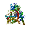 3k1oSC 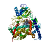 3kswC S: Starting model for refinement C: citing same article ( |
|---|---|
| Similar structure data |
-Links
 PDBj
PDBj
- Assembly
| Deposited unit | 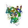
| ||||||||
|---|---|---|---|---|---|---|---|---|---|
| 1 |
| ||||||||
| Unit cell |
|
-Components
| #1: Protein | Mass: 52925.340 Da / Num. of mol.: 1 / Fragment: UNP RESIDUES 24-481 / Mutation: F24K, N25T, T26S, T27G, R28K, P29G, T30K, D31L Source method: isolated from a genetically manipulated source Source: (gene. exp.)  References: UniProt: Q5I4E1, UniProt: Q7Z1V1*PLUS, sterol 14alpha-demethylase |
|---|---|
| #2: Chemical | ChemComp-HEM /   Mass: 616.487 Da / Num. of mol.: 1 / Source method: obtained synthetically / Formula: C34H32FeN4O4 Mass: 616.487 Da / Num. of mol.: 1 / Source method: obtained synthetically / Formula: C34H32FeN4O4 |
| #3: Chemical | ChemComp-TPF /   Mass: 306.271 Da / Num. of mol.: 1 / Source method: obtained synthetically / Formula: C13H12F2N6O / Comment: medication*YM Mass: 306.271 Da / Num. of mol.: 1 / Source method: obtained synthetically / Formula: C13H12F2N6O / Comment: medication*YM |
-Experimental details
-Experiment
| Experiment | Method: X-RAY DIFFRACTION / Number of used crystals: 1 |
|---|
- Sample preparation
| Crystal | Density Matthews: 2.37 Å3/Da / Density % sol: 48.2 % |
|---|---|
| Crystal grow | Temperature: 298 K / Method: vapor diffusion, hanging drop / pH: 7.2 Details: PEG 3350, POTASSIUM FORMATE, SODIUM CHLORIDE, pH 7.2, VAPOR DIFFUSION, HANGING DROP, temperature 298K |
-Data collection
| Diffraction | Mean temperature: 100 K |
|---|---|
| Diffraction source | Source: SYNCHROTRON / Site: APS  / Beamline: 22-ID / Wavelength: 0.97928 Å / Beamline: 22-ID / Wavelength: 0.97928 Å |
| Detector | Type: MARMOSAIC 300 mm CCD / Detector: CCD / Date: Dec 14, 2005 |
| Radiation | Monochromator: DIAMOND / Protocol: SINGLE WAVELENGTH / Monochromatic (M) / Laue (L): M / Scattering type: x-ray |
| Radiation wavelength | Wavelength: 0.97928 Å / Relative weight: 1 |
| Reflection | Resolution: 2.85→50 Å / Num. all: 12689 / Num. obs: 12181 / % possible obs: 96 % / Observed criterion σ(F): 1.4 / Observed criterion σ(I): 1.4 / Redundancy: 3.6 % / Biso Wilson estimate: 48.4 Å2 / Rmerge(I) obs: 0.06 / Net I/σ(I): 24.8 |
| Reflection shell | Resolution: 2.85→2.95 Å / Redundancy: 3.3 % / Rmerge(I) obs: 0.502 / Mean I/σ(I) obs: 1.4 / Num. unique all: 2136 / % possible all: 95.9 |
- Processing
| Software |
| |||||||||||||||||||||||||||||||||||||||||||||||||||||||||||||||||
|---|---|---|---|---|---|---|---|---|---|---|---|---|---|---|---|---|---|---|---|---|---|---|---|---|---|---|---|---|---|---|---|---|---|---|---|---|---|---|---|---|---|---|---|---|---|---|---|---|---|---|---|---|---|---|---|---|---|---|---|---|---|---|---|---|---|---|
| Refinement | Method to determine structure: MOLECULAR REPLACEMENT Starting model: PDB entry 3K1O Resolution: 2.85→28.83 Å / Cor.coef. Fo:Fc: 0.943 / Cor.coef. Fo:Fc free: 0.899 / SU B: 46.601 / SU ML: 0.396 / TLS residual ADP flag: LIKELY RESIDUAL / Cross valid method: THROUGHOUT / σ(F): 1.4 / ESU R Free: 0.47 / Stereochemistry target values: MAXIMUM LIKELIHOOD / Details: HYDROGENS HAVE BEEN ADDED IN THE RIDING POSITIONS
| |||||||||||||||||||||||||||||||||||||||||||||||||||||||||||||||||
| Solvent computation | Ion probe radii: 0.8 Å / Shrinkage radii: 0.8 Å / VDW probe radii: 1.4 Å / Solvent model: MASK | |||||||||||||||||||||||||||||||||||||||||||||||||||||||||||||||||
| Displacement parameters | Biso mean: 43.49 Å2
| |||||||||||||||||||||||||||||||||||||||||||||||||||||||||||||||||
| Refinement step | Cycle: LAST / Resolution: 2.85→28.83 Å
| |||||||||||||||||||||||||||||||||||||||||||||||||||||||||||||||||
| Refine LS restraints |
| |||||||||||||||||||||||||||||||||||||||||||||||||||||||||||||||||
| LS refinement shell | Resolution: 2.85→2.924 Å / Total num. of bins used: 20
| |||||||||||||||||||||||||||||||||||||||||||||||||||||||||||||||||
| Refinement TLS params. | Method: refined / Origin x: 9.732 Å / Origin y: -24.882 Å / Origin z: 18.256 Å
|
