 Movie
Movie Controller

[English] 日本語
 Yorodumi
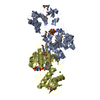
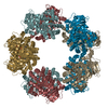
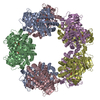
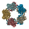
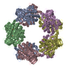
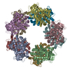
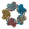
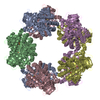
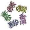
- PDB-3kdn: Crystal structure of Type III Rubisco SP4 mutant complexed with 2-CABP -
+ Open data

- Basic information
| Entry | Database: PDB / ID: 3kdn | ||||||
|---|---|---|---|---|---|---|---|
| Title | Crystal structure of Type III Rubisco SP4 mutant complexed with 2-CABP | ||||||
 Components Components | Ribulose bisphosphate carboxylase | ||||||
 Keywords Keywords | LYASE / ribulose-1 / 5-bisphosphate carboxylase/oxygenase / Rubisco / Carbon dioxide fixation / Magnesium / Metal-binding / Monooxygenase | ||||||
| Function / homology |  Function and homology information Function and homology informationAMP catabolic process / ribulose-bisphosphate carboxylase / ribulose-bisphosphate carboxylase activity / carbon fixation / oxidoreductase activity / magnesium ion binding Similarity search - Function | ||||||
| Biological species |   Thermococcus kodakaraensis (archaea) Thermococcus kodakaraensis (archaea) | ||||||
| Method |  X-RAY DIFFRACTION / SYNCHROTRON / MOLECULAR REPLACEMENT / Resolution: 2.09 Å X-RAY DIFFRACTION / SYNCHROTRON / MOLECULAR REPLACEMENT / Resolution: 2.09 Å | ||||||
 Authors Authors | Nishitani, Y. / Fujihashi, M. / Doi, T. / Yoshida, S. / Atomi, H. / Imanaka, T. / Miki, K. | ||||||
 Citation Citation | Journal: J.Biol.Chem. / Year: 2010 Title: Structure-based catalytic optimization of a type III Rubisco from a hyperthermophile Authors: Nishitani, Y. / Yoshida, S. / Fujihashi, M. / Kitagawa, K. / Doi, T. / Atomi, H. / Imanaka, T. / Miki, K. | ||||||
| History |
|
- Structure visualization
| Structure viewer | Molecule: MolmilJmol/JSmol |
|---|
- Downloads & links
-Download
| PDBx/mmCIF format | 3kdn.cif.gz | 884.1 KB | Display | PDBx/mmCIF format |
|---|---|---|---|---|
| PDB format | pdb3kdn.ent.gz | 727.8 KB | Display | PDB format |
| PDBx/mmJSON format | 3kdn.json.gz | Tree view | PDBx/mmJSON format | |
| Others |  Other downloads Other downloads |
-Validation report
| Summary document | 3kdn_validation.pdf.gz | 3.5 MB | Display | wwPDB validaton report |
|---|---|---|---|---|
| Full document | 3kdn_full_validation.pdf.gz | 3.6 MB | Display | |
| Data in XML | 3kdn_validation.xml.gz | 187 KB | Display | |
| Data in CIF | 3kdn_validation.cif.gz | 252.1 KB | Display | |
| Arichive directory | https://data.pdbj.org/pub/pdb/validation_reports/kd/3kdnftp://data.pdbj.org/pub/pdb/validation_reports/kd/3kdn | HTTPS FTP |
-Related structure data
| Related structure data |  3a12C  3kdoC  1gehS S: Starting model for refinement C: citing same article ( |
|---|---|
| Similar structure data |

-Links
 PDBj
PDBj
- Assembly
| Deposited unit | 
| ||||||||
|---|---|---|---|---|---|---|---|---|---|
| 1 |
| ||||||||
| Unit cell |
|
-Components
| #1: Protein | Mass: 49848.477 Da / Num. of mol.: 10 / Mutation: G326E, K327R, W328D, D329I, V330T Source method: isolated from a genetically manipulated source Source: (gene. exp.) Thermococcus kodakaraensis (archaea) / Strain: KOD1 / Gene: rbcL, TK2290 / Plasmid: pET-21a(+) / Production host:  References: UniProt: O93627, ribulose-bisphosphate carboxylase #2: Chemical | ChemComp-MG /   Mass: 24.305 Da / Num. of mol.: 10 / Source method: obtained synthetically / Formula: Mg Mass: 24.305 Da / Num. of mol.: 10 / Source method: obtained synthetically / Formula: Mg#3: Sugar | ChemComp-CAP /   Type: saccharide / Mass: 356.115 Da / Num. of mol.: 10 / Source method: obtained synthetically / Formula: C6H14O13P2 Type: saccharide / Mass: 356.115 Da / Num. of mol.: 10 / Source method: obtained synthetically / Formula: C6H14O13P2#4: Water | ChemComp-HOH / |  Mass: 18.015 Da / Num. of mol.: 2652 / Source method: isolated from a natural source / Formula: H2O Mass: 18.015 Da / Num. of mol.: 2652 / Source method: isolated from a natural source / Formula: H2O |
|---|
-Experimental details
-Experiment
| Experiment | Method: X-RAY DIFFRACTION / Number of used crystals: 1 |
|---|
- Sample preparation
| Crystal | Density Matthews: 3.11 Å3/Da / Density % sol: 60.42 % |
|---|---|
| Crystal grow | Temperature: 293 K / Method: vapor diffusion, hanging drop / pH: 6 Details: 0.1M Acetate, 80mM CaCl2, 6% PEG6000, 10% MPD, pH6.0, VAPOR DIFFUSION, HANGING DROP, temperature 293K |
-Data collection
| Diffraction | Mean temperature: 100 K |
|---|---|
| Diffraction source | Source: SYNCHROTRON / Site: SPring-8  / Beamline: BL41XU / Wavelength: 1 Å / Beamline: BL41XU / Wavelength: 1 Å |
| Detector | Type: RAYONIX MX225HE / Detector: CCD / Date: Jul 3, 2009 |
| Radiation | Protocol: SINGLE WAVELENGTH / Monochromatic (M) / Laue (L): M / Scattering type: x-ray |
| Radiation wavelength | Wavelength: 1 Å / Relative weight: 1 |
| Reflection | Resolution: 2.09→50 Å / Num. obs: 345208 / % possible obs: 96.5 % / Rsym value: 0.071 / Net I/σ(I): 15.4 |
| Reflection shell | Resolution: 2.09→2.13 Å / Mean I/σ(I) obs: 2.3 / Rsym value: 0.384 / % possible all: 70.3 |
- Processing
| Software |
| |||||||||||||||||||||||||||||||||||||||||||||||||||||||||||||||||
|---|---|---|---|---|---|---|---|---|---|---|---|---|---|---|---|---|---|---|---|---|---|---|---|---|---|---|---|---|---|---|---|---|---|---|---|---|---|---|---|---|---|---|---|---|---|---|---|---|---|---|---|---|---|---|---|---|---|---|---|---|---|---|---|---|---|---|
| Refinement | Method to determine structure: MOLECULAR REPLACEMENT Starting model: PDB ENTRY 1GEH Resolution: 2.09→42.86 Å / Cor.coef. Fo:Fc: 0.933 / Cor.coef. Fo:Fc free: 0.905 / SU B: 4.808 / SU ML: 0.128 / Cross valid method: THROUGHOUT / ESU R: 0.208 / ESU R Free: 0.183 / Stereochemistry target values: MAXIMUM LIKELIHOOD / Details: HYDROGENS HAVE BEEN ADDED IN THE RIDING POSITIONS
| |||||||||||||||||||||||||||||||||||||||||||||||||||||||||||||||||
| Solvent computation | Ion probe radii: 0.8 Å / Shrinkage radii: 0.8 Å / VDW probe radii: 1.2 Å / Solvent model: MASK | |||||||||||||||||||||||||||||||||||||||||||||||||||||||||||||||||
| Displacement parameters | Biso mean: 26.249 Å2
| |||||||||||||||||||||||||||||||||||||||||||||||||||||||||||||||||
| Refinement step | Cycle: LAST / Resolution: 2.09→42.86 Å
| |||||||||||||||||||||||||||||||||||||||||||||||||||||||||||||||||
| Refine LS restraints |
| |||||||||||||||||||||||||||||||||||||||||||||||||||||||||||||||||
| LS refinement shell | Resolution: 2.09→2.144 Å / Total num. of bins used: 20
|
