 Movie
Movie Controller

[English] 日本語
 Yorodumi
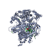
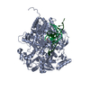
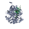
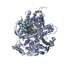
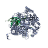
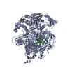
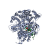
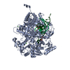
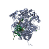
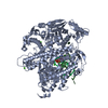
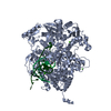
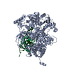
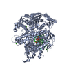
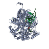
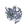
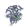
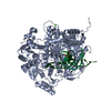
- PDB-3kd1: Closed binary complex of an RB69 gp43 fingers domain mutant compl... -
+ Open data

- Basic information
| Entry | Database: PDB / ID: 3kd1 | ||||||
|---|---|---|---|---|---|---|---|
| Title | Closed binary complex of an RB69 gp43 fingers domain mutant complexed with an acyclic GMP terminated primer template pair. | ||||||
 Components Components |
| ||||||
 Keywords Keywords | TRANSFERASE/DNA / gp43 / polymerase / HCMV / human cytomegalovirus / acyclovir / acyclic guanosine / foscavir / foscarnet / phosphonoformic acid / DNA replication / DNA-binding / DNA-directed DNA polymerase / Exonuclease / Hydrolase / Nuclease / Nucleotidyltransferase / Transferase / TRANSFERASE-DNA COMPLEX | ||||||
| Function / homology |  Function and homology information Function and homology informationbidirectional double-stranded viral DNA replication / Hydrolases; Acting on ester bonds; Exodeoxyribonucleases producing 5'-phosphomonoesters / 3'-5' exonuclease activity / DNA-templated DNA replication / DNA-directed DNA polymerase / DNA-directed DNA polymerase activity / nucleotide binding / DNA binding / metal ion binding Similarity search - Function | ||||||
| Biological species |  Enterobacteria phage RB69 (virus) Enterobacteria phage RB69 (virus) | ||||||
| Method |  X-RAY DIFFRACTION / SYNCHROTRON / isomorphous replacement / Resolution: 2.66 Å X-RAY DIFFRACTION / SYNCHROTRON / isomorphous replacement / Resolution: 2.66 Å | ||||||
 Authors Authors | Zahn, K.E. / Doublie, S. | ||||||
 Citation Citation | Journal: J.Biol.Chem. / Year: 2011 Title: Phosphonoformic acid inhibits viral replication by trapping the closed form of the DNA polymerase. Authors: Zahn, K.E. / Tchesnokov, E.P. / Gotte, M. / Doublie, S. | ||||||
| History |
|
- Structure visualization
| Structure viewer | Molecule: MolmilJmol/JSmol |
|---|
- Downloads & links
-Download
| PDBx/mmCIF format | 3kd1.cif.gz | 219.1 KB | Display | PDBx/mmCIF format |
|---|---|---|---|---|
| PDB format | pdb3kd1.ent.gz | 168.3 KB | Display | PDB format |
| PDBx/mmJSON format | 3kd1.json.gz | Tree view | PDBx/mmJSON format | |
| Others |  Other downloads Other downloads |
-Validation report
| Arichive directory | https://data.pdbj.org/pub/pdb/validation_reports/kd/3kd1ftp://data.pdbj.org/pub/pdb/validation_reports/kd/3kd1 | HTTPS FTP |
|---|
-Related structure data
| Related structure data | 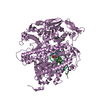 3kd5C 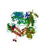 1ih7S C: citing same article ( S: Starting model for refinement |
|---|---|
| Similar structure data |


-Links
 PDBj
PDBj


- Assembly
| Deposited unit | 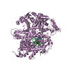
| ||||||||
|---|---|---|---|---|---|---|---|---|---|
| 1 |
| ||||||||
| Unit cell |
|
-Components
| #1: DNA chain | Mass: 5492.554 Da / Num. of mol.: 1 / Source method: obtained synthetically / Details: Oligonucleotide was chemically synthesized |
|---|---|
| #2: DNA chain | Mass: 4278.780 Da / Num. of mol.: 1 / Fragment: Acyclic GMP terminated primer DNA / Source method: obtained synthetically / Details: Oligonucleotide was chemically synthesized |
| #3: Protein | Mass: 105806.359 Da / Num. of mol.: 1 Fragment: RB69 gp43 exo- chimera containing elements from the fingers domain of the human cytomegalovirus DNA polymerase. Mutation: D222A V478W F479V N480S I557M N558A R559L L561V I562T I563C Source method: isolated from a genetically manipulated source Source: (gene. exp.) Enterobacteria phage RB69 (virus) / Gene: 43, RB69 gp43 / Plasmid: pPR-IBA1 / Production host:  |
| #4: Chemical | ChemComp-MG /   Mass: 24.305 Da / Num. of mol.: 1 / Source method: obtained synthetically / Formula: Mg Mass: 24.305 Da / Num. of mol.: 1 / Source method: obtained synthetically / Formula: Mg |
| #5: Water | ChemComp-HOH /  Mass: 18.015 Da / Num. of mol.: 321 / Source method: isolated from a natural source / Formula: H2O Mass: 18.015 Da / Num. of mol.: 321 / Source method: isolated from a natural source / Formula: H2O |
-Experimental details
-Experiment
| Experiment | Method: X-RAY DIFFRACTION / Number of used crystals: 1 |
|---|
- Sample preparation
| Crystal | Density Matthews: 2.72 Å3/Da / Density % sol: 54.71 % |
|---|---|
| Crystal grow | Temperature: 298 K / Method: vapor diffusion, hanging drop / pH: 7.5 Details: 5% PEG 20000, 0.1M sodium acetate pH 5, 0.1M magnesium acetate, 0.1M Tris-HCl pH 7.5, VAPOR DIFFUSION, HANGING DROP, temperature 298K |
-Data collection
| Diffraction | Mean temperature: 200 K |
|---|---|
| Diffraction source | Source: SYNCHROTRON / Site: APS  / Beamline: 23-ID-B / Wavelength: 1.03315 Å / Beamline: 23-ID-B / Wavelength: 1.03315 Å |
| Detector | Type: MAR scanner 300 mm plate / Detector: IMAGE PLATE / Date: Aug 15, 2009 Details: Si(111) Double Crystal Monochrometer. Adjustable focusing mirrors in K-B geomet ry |
| Radiation | Monochromator: Double crystal cryo-cooled Si(111) / Protocol: SINGLE WAVELENGTH / Monochromatic (M) / Laue (L): M / Scattering type: x-ray |
| Radiation wavelength | Wavelength: 1.03315 Å / Relative weight: 1 |
| Reflection | Resolution: 2.66→50 Å / Num. all: 36963 / Num. obs: 36963 / % possible obs: 100 % / Observed criterion σ(F): 0 / Observed criterion σ(I): 0 / Redundancy: 7.6 % / Rmerge(I) obs: 0.106 / Net I/σ(I): 17.7 |
| Reflection shell | Resolution: 2.66→2.76 Å / Redundancy: 7.2 % / Mean I/σ(I) obs: 2.24 / % possible all: 100 |
- Processing
| Software |
| ||||||||||||||||||||||||||||||||||||||||||||||||||||||||||||||||||||||||||||||||||||||||||||||||||||||||||||||||||||||||||||||||||||||||||||||||||||||||||||||||||||||||||||||||||||||||||||||||||||||||
|---|---|---|---|---|---|---|---|---|---|---|---|---|---|---|---|---|---|---|---|---|---|---|---|---|---|---|---|---|---|---|---|---|---|---|---|---|---|---|---|---|---|---|---|---|---|---|---|---|---|---|---|---|---|---|---|---|---|---|---|---|---|---|---|---|---|---|---|---|---|---|---|---|---|---|---|---|---|---|---|---|---|---|---|---|---|---|---|---|---|---|---|---|---|---|---|---|---|---|---|---|---|---|---|---|---|---|---|---|---|---|---|---|---|---|---|---|---|---|---|---|---|---|---|---|---|---|---|---|---|---|---|---|---|---|---|---|---|---|---|---|---|---|---|---|---|---|---|---|---|---|---|---|---|---|---|---|---|---|---|---|---|---|---|---|---|---|---|---|---|---|---|---|---|---|---|---|---|---|---|---|---|---|---|---|---|---|---|---|---|---|---|---|---|---|---|---|---|---|---|---|---|
| Refinement | Method to determine structure: isomorphous replacement Starting model: PDB entry 1IH7 Resolution: 2.66→30 Å / Cor.coef. Fo:Fc: 0.947 / Cor.coef. Fo:Fc free: 0.919 / SU B: 22.55 / SU ML: 0.249 / Isotropic thermal model: ISOTROPIC / Cross valid method: THROUGHOUT / ESU R Free: 0.342 / Stereochemistry target values: MAXIMUM LIKELIHOOD
| ||||||||||||||||||||||||||||||||||||||||||||||||||||||||||||||||||||||||||||||||||||||||||||||||||||||||||||||||||||||||||||||||||||||||||||||||||||||||||||||||||||||||||||||||||||||||||||||||||||||||
| Solvent computation | Ion probe radii: 0.8 Å / Shrinkage radii: 0.8 Å / VDW probe radii: 1.4 Å / Solvent model: BABINET MODEL WITH MASK | ||||||||||||||||||||||||||||||||||||||||||||||||||||||||||||||||||||||||||||||||||||||||||||||||||||||||||||||||||||||||||||||||||||||||||||||||||||||||||||||||||||||||||||||||||||||||||||||||||||||||
| Displacement parameters | Biso mean: 41.056 Å2
| ||||||||||||||||||||||||||||||||||||||||||||||||||||||||||||||||||||||||||||||||||||||||||||||||||||||||||||||||||||||||||||||||||||||||||||||||||||||||||||||||||||||||||||||||||||||||||||||||||||||||
| Refinement step | Cycle: LAST / Resolution: 2.66→30 Å
| ||||||||||||||||||||||||||||||||||||||||||||||||||||||||||||||||||||||||||||||||||||||||||||||||||||||||||||||||||||||||||||||||||||||||||||||||||||||||||||||||||||||||||||||||||||||||||||||||||||||||
| Refine LS restraints |
| ||||||||||||||||||||||||||||||||||||||||||||||||||||||||||||||||||||||||||||||||||||||||||||||||||||||||||||||||||||||||||||||||||||||||||||||||||||||||||||||||||||||||||||||||||||||||||||||||||||||||
| LS refinement shell | Resolution: 2.66→2.728 Å / Total num. of bins used: 20
| ||||||||||||||||||||||||||||||||||||||||||||||||||||||||||||||||||||||||||||||||||||||||||||||||||||||||||||||||||||||||||||||||||||||||||||||||||||||||||||||||||||||||||||||||||||||||||||||||||||||||
| Refinement TLS params. | Method: refined / Refine-ID: X-RAY DIFFRACTION
| ||||||||||||||||||||||||||||||||||||||||||||||||||||||||||||||||||||||||||||||||||||||||||||||||||||||||||||||||||||||||||||||||||||||||||||||||||||||||||||||||||||||||||||||||||||||||||||||||||||||||
| Refinement TLS group |
|
