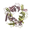
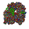
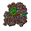
CRYSTAL PACKING AND ANALYTICAL SIZE EXCLUSION CHROMATOGRAPHY ANALYSIS SUPPORT THE ASSIGNMENT OF A TRIMER AS A SIGNIFICANT OLIGOMERIZATION STATE IN SOLUTION.
-
Components
-
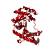
Protein , 1 types, 1 molecules A
#1: Protein
phagerelatedexonuclease
Mass: 25887.744 Da / Num. of mol.: 1 Source method: isolated from a genetically manipulated source Source: (gene. exp.) Haemophilus somnus 129PT (bacteria) / Strain: 129Pt / Gene: HS_1420 / Plasmid: SpeedET / Production host: Escherichia Coli (E. coli) / Strain (production host): HK100 / References: UniProt: Q0I4G3
Mass: 18.015 Da / Num. of mol.: 122 / Source method: isolated from a natural source / Formula: H2O
-
Details
Has protein modification
Y
Sequence details
THIS CONSTRUCT (1-222) WAS EXPRESSED WITH THE PURIFICATION TAG MGSDKIHHHHHHENLYFQG. THE TAG WAS ...THIS CONSTRUCT (1-222) WAS EXPRESSED WITH THE PURIFICATION TAG MGSDKIHHHHHHENLYFQG. THE TAG WAS REMOVED WITH TEV PROTEASE LEAVING ONLY A GLYCINE (0) FOLLOWED BY THE TARGET SEQUENCE.
-
Experimental details
-
Experiment
Experiment
Method: X-RAY DIFFRACTION / Number of used crystals: 1
-
Sample preparation
Crystal
Density Matthews: 3.03 Å3/Da / Density % sol: 59.41 %
Crystal grow
Temperature: 277 K / Method: vapor diffusion, sitting drop / pH: 6.5 Details: 10.0000% Glycerol, 1.0000M NaCl, 30.0000% PEG-600, 0.1M Cacodylate pH 6.5, NANODROP, VAPOR DIFFUSION, SITTING DROP, temperature 277K
Monochromator: Single crystal Si(111) bent monochromator (horizontal focusing) Protocol: MAD / Monochromatic (M) / Laue (L): M / Scattering type: x-ray
Radiation wavelength
ID
Wavelength (Å)
Relative weight
1
0.91837
1
2
0.97947
1
Reflection
Resolution: 2.15→28.341 Å / Num. obs: 17252 / % possible obs: 100 % / Redundancy: 7.4 % / Biso Wilson estimate: 32.941 Å2 / Rmerge(I) obs: 0.129 / Rsym value: 0.129 / Net I/σ(I): 12.8
Reflection shell
Diffraction-ID: 1
Resolution (Å)
Redundancy (%)
Rmerge(I) obs
Mean I/σ(I) obs
Num. measured all
Num. unique all
Rsym value
% possible all
2.15-2.21
7.5
0.836
2.1
9647
1285
0.836
100
2.21-2.27
7.5
0.682
1.1
9033
1205
0.682
100
2.27-2.33
7.5
0.575
1.3
9065
1204
0.575
100
2.33-2.4
7.5
0.495
1.5
8654
1156
0.495
100
2.4-2.48
7.5
0.399
1.9
8612
1145
0.399
100
2.48-2.57
7.5
0.333
2.3
8152
1083
0.333
100
2.57-2.67
7.5
0.313
2.4
7814
1048
0.313
100
2.67-2.78
7.6
0.244
3.1
7670
1015
0.244
100
2.78-2.9
7.5
0.204
3.7
7268
975
0.204
100
2.9-3.04
7.5
0.157
4.7
7057
941
0.157
100
3.04-3.21
7.5
0.128
5.6
6641
884
0.128
100
3.21-3.4
7.5
0.107
6.4
6303
844
0.107
100
3.4-3.63
7.4
0.092
7.2
5873
794
0.092
100
3.63-3.93
7.4
0.081
7.7
5526
749
0.081
100
3.93-4.3
7.4
0.071
9.2
5039
683
0.071
100
4.3-4.81
7.4
0.066
9.7
4631
630
0.066
100
4.81-5.55
7.3
0.07
9.2
4000
546
0.07
100
5.55-6.8
7.2
0.073
9.3
3404
476
0.073
100
6.8-9.62
7.1
0.061
9.8
2651
375
0.061
100
9.62-28.34
6.4
0.055
11.5
1365
214
0.055
96.6
-
Phasing
Phasing
Method: MAD
-
Processing
Software
Name
Version
Classification
NB
REFMAC
5.5.0053
refinement
PHENIX
refinement
SOLVE
phasing
MolProbity
3beta29
modelbuilding
SCALA
3.2.5
datascaling
PDB_EXTRACT
3.006
dataextraction
MOSFLM
datareduction
Refinement
Method to determine structure: MAD / Resolution: 2.15→28.341 Å / Cor.coef. Fo:Fc: 0.961 / Cor.coef. Fo:Fc free: 0.939 / Occupancy max: 1 / Occupancy min: 0.4 / SU B: 9.253 / SU ML: 0.107 / TLS residual ADP flag: LIKELY RESIDUAL / Cross valid method: THROUGHOUT / σ(F): 0 / ESU R: 0.18 / ESU R Free: 0.16 Stereochemistry target values: MAXIMUM LIKELIHOOD WITH PHASES Details: 1. HYDROGENS HAVE BEEN ADDED IN THE RIDING POSITIONS. 2. ATOM RECORD CONTAINS RESIDUAL B FACTORS ONLY. 3. A MET-INHIBITION PROTOCOL WAS USED FOR SELENOMETHIONINE INCORPORATION DURING PROTEIN ...Details: 1. HYDROGENS HAVE BEEN ADDED IN THE RIDING POSITIONS. 2. ATOM RECORD CONTAINS RESIDUAL B FACTORS ONLY. 3. A MET-INHIBITION PROTOCOL WAS USED FOR SELENOMETHIONINE INCORPORATION DURING PROTEIN EXPRESSION. THE OCCUPANCY OF THE SE ATOMS IN THE MSE RESIDUES WAS REDUCED TO 0.75 TO ACCOUNT FOR THE REDUCED SCATTERING POWER DUE TO PARTIAL S-MET INCORPORATION. 4. SODIUM (NA) AND CHLORIDE (CL) IONS, GLYCEROL (GOL), AND POLYETHYLENE GLYCOL (PEG) FROM THE CRYSTALLIZATION SOLUTION HAVE BEEN MODELED INTO THE SOLVENT STRUCTURE. 5. TLS GROUPS WERE ASSIGNED WITH THE AID OF THE TLSMD SERVER.
Rfactor
Num. reflection
% reflection
Selection details
Rfree
0.212
874
5.1 %
RANDOM
Rwork
0.172
-
-
-
obs
0.174
17249
99.99 %
-
Solvent computation
Ion probe radii: 0.8 Å / Shrinkage radii: 0.8 Å / VDW probe radii: 1.4 Å / Solvent model: MASK
In the structure databanks used in Yorodumi, some data are registered as the other names, "COVID-19 virus" and "2019-nCoV". Here are the details of the virus and the list of structure data.
Jan 31, 2019. EMDB accession codes are about to change! (news from PDBe EMDB page)
EMDB accession codes are about to change! (news from PDBe EMDB page)
The allocation of 4 digits for EMDB accession codes will soon come to an end. Whilst these codes will remain in use, new EMDB accession codes will include an additional digit and will expand incrementally as the available range of codes is exhausted. The current 4-digit format prefixed with “EMD-” (i.e. EMD-XXXX) will advance to a 5-digit format (i.e. EMD-XXXXX), and so on. It is currently estimated that the 4-digit codes will be depleted around Spring 2019, at which point the 5-digit format will come into force.
The EM Navigator/Yorodumi systems omit the EMD- prefix.
Related info.:Q: What is EMD? / ID/Accession-code notation in Yorodumi/EM Navigator
Yorodumi is a browser for structure data from EMDB, PDB, SASBDB, etc.
This page is also the successor to EM Navigator detail page, and also detail information page/front-end page for Omokage search.
The word "yorodu" (or yorozu) is an old Japanese word meaning "ten thousand". "mi" (miru) is to see.
Related info.:EMDB / PDB / SASBDB / Comparison of 3 databanks / Yorodumi Search / Aug 31, 2016. New EM Navigator & Yorodumi / Yorodumi Papers / Jmol/JSmol / Function and homology information / Changes in new EM Navigator and Yorodumi
 Movie
Movie Controller

 Yorodumi
 Open data
 Basic information
 Components
 Keywords
 Function and homology information
 Haemophilus somnus 129PT (bacteria)
 X-RAY DIFFRACTION /
  Authors
 Citation
 Structure visualization
 Downloads & links
 Other downloads
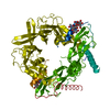


 PDBj
PDBj


 Assembly
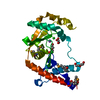


 Mass: 22.990 Da / Num. of mol.: 1 / Source method: obtained synthetically / Formula: Na
 Mass: 35.453 Da / Num. of mol.: 2 / Source method: obtained synthetically / Formula: Cl
 Mass: 92.094 Da / Num. of mol.: 1 / Source method: obtained synthetically / Formula: C3H8O3
 Mass: 106.120 Da / Num. of mol.: 2 / Source method: obtained synthetically / Formula: C4H10O3
 Sample preparation
 / Beamline: BL11-1 / Wavelength: 0.91837,0.97947
 Processing