 Movie
Movie Controller


+ Open data

- Basic information
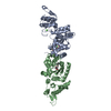
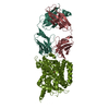
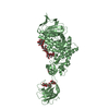
| Entry | Database: PDB / ID: 3k8m | |||||||||
|---|---|---|---|---|---|---|---|---|---|---|
| Title | Crystal structure of SusG with acarbose | |||||||||
 Components Components | Alpha-amylase, susG | |||||||||
 Keywords Keywords | MEMBRANE PROTEIN / amylase / alpha8/beta8 barrel / CBM / beta-sandwich | |||||||||
| Function / homology |  Function and homology information Function and homology informationstarch catabolic process / starch binding / alpha-amylase / outer membrane / alpha-amylase activity / oligosaccharide catabolic process / cell outer membrane / calcium ion binding / magnesium ion binding Similarity search - Function | |||||||||
| Biological species |  Bacteroides thetaiotaomicron (bacteria) Bacteroides thetaiotaomicron (bacteria) | |||||||||
| Method |  X-RAY DIFFRACTION / SYNCHROTRON / MOLECULAR REPLACEMENT / molecular replacement / Resolution: 2.5 Å X-RAY DIFFRACTION / SYNCHROTRON / MOLECULAR REPLACEMENT / molecular replacement / Resolution: 2.5 Å | |||||||||
 Authors Authors | Koropatkin, N.M. / Smith, T.J. | |||||||||
 Citation Citation | Journal: Structure / Year: 2010 Title: SusG: A Unique Cell-Membrane-Associated alpha-Amylase from a Prominent Human Gut Symbiont Targets Complex Starch Molecules. Authors: Koropatkin, N.M. / Smith, T.J. | |||||||||
| History |
|
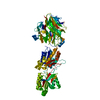
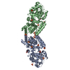
- Structure visualization
| Structure viewer | Molecule: MolmilJmol/JSmol |
|---|
- Downloads & links
-Download
| PDBx/mmCIF format | 3k8m.cif.gz | 277.2 KB | Display | PDBx/mmCIF format |
|---|---|---|---|---|
| PDB format | pdb3k8m.ent.gz | 223 KB | Display | PDB format |
| PDBx/mmJSON format | 3k8m.json.gz | Tree view | PDBx/mmJSON format | |
| Others |  Other downloads Other downloads |
-Validation report
| Arichive directory | https://data.pdbj.org/pub/pdb/validation_reports/k8/3k8mftp://data.pdbj.org/pub/pdb/validation_reports/k8/3k8m | HTTPS FTP |
|---|
-Related structure data
| Related structure data | 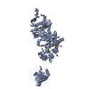 3k8kSC 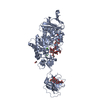 3k8lC S: Starting model for refinement C: citing same article ( |
|---|---|
| Similar structure data |


-Links
 PDBj
PDBj


- Assembly
| Deposited unit | 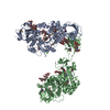
| ||||||||
|---|---|---|---|---|---|---|---|---|---|
| 1 | 
| ||||||||
| 2 | 
| ||||||||
| Unit cell |
|
-Components
-Protein , 1 types, 2 molecules AB
| #1: Protein | Mass: 75827.727 Da / Num. of mol.: 2 Source method: isolated from a genetically manipulated source Source: (gene. exp.) Bacteroides thetaiotaomicron (bacteria)Strain: VPI-5482 / Gene: BT_3698, SusG / Plasmid: pET-28rTEV / Production host: |
|---|
-Sugars , 3 types, 8 molecules
| #2: Polysaccharide | 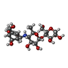  Type: oligosaccharide, Oligosaccharide / Class: Inhibitor / Mass: 483.465 Da / Num. of mol.: 2 Type: oligosaccharide, Oligosaccharide / Class: Inhibitor / Mass: 483.465 Da / Num. of mol.: 2Source method: isolated from a genetically manipulated source Details: oligosaccharide / References: acarbose-derived trisaccharide #3: Polysaccharide | 4,6-dideoxy-4-{[(1S,4R,5S,6S)-4,5,6-trihydroxy-3-(hydroxymethyl)cyclohex-2-en-1-yl]amino}-alpha-D- ...4,6-dideoxy-4-{[(1S,4R,5S,6S)-4,5,6-trihydroxy-3-(hydroxymethyl)cyclohex-2-en-1-yl]amino}-alpha-D-glucopyranose-(1-4)-alpha-D-glucopyranose-(1-4)-alpha-D-glucopyranose / alpha-acarbose 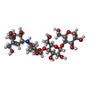  Type: oligosaccharide, Oligosaccharide / Class: Inhibitor / Mass: 645.606 Da / Num. of mol.: 4 Type: oligosaccharide, Oligosaccharide / Class: Inhibitor / Mass: 645.606 Da / Num. of mol.: 4Source method: isolated from a genetically manipulated source Details: oligosaccharide / References: alpha-acarbose #4: Polysaccharide |   Source method: isolated from a genetically manipulated source Details: oligosaccharide / References: alpha-maltose |
|---|
-Non-polymers , 3 types, 175 molecules 


| #5: Chemical | ChemComp-CA /  Mass: 40.078 Da / Num. of mol.: 4 / Source method: obtained synthetically / Formula: Ca Mass: 40.078 Da / Num. of mol.: 4 / Source method: obtained synthetically / Formula: Ca#6: Chemical | ChemComp-EDO / |  Mass: 62.068 Da / Num. of mol.: 1 / Source method: obtained synthetically / Formula: C2H6O2 Mass: 62.068 Da / Num. of mol.: 1 / Source method: obtained synthetically / Formula: C2H6O2#7: Water | ChemComp-HOH / | Mass: 18.015 Da / Num. of mol.: 170 / Source method: isolated from a natural source / Formula: H2O |
|---|
-Details
| Has protein modification | Y |
|---|
-Experimental details
-Experiment
| Experiment | Method: X-RAY DIFFRACTION / Number of used crystals: 1 |
|---|
- Sample preparation
| Crystal | Density Matthews: 3.44 Å3/Da / Density % sol: 64.25 % |
|---|---|
| Crystal grow | Temperature: 298 K / Method: vapor diffusion, hanging drop / pH: 7.5 Details: 22% PEG 4000, 50 mM LiSO4, 100mM HEPES, 10mM acarbose, 0.5mM CaCl2, pH 7.5, VAPOR DIFFUSION, HANGING DROP, temperature 298K |
-Data collection
| Diffraction | Mean temperature: 100 K |
|---|---|
| Diffraction source | Source: SYNCHROTRON / Site: APS  / Beamline: 19-ID / Wavelength: 0.97929 Å / Beamline: 19-ID / Wavelength: 0.97929 Å |
| Detector | Type: SBC-3 / Detector: CCD / Date: Jan 1, 2008 / Details: mirrors |
| Radiation | Monochromator: Si 111 / Protocol: MAD / Monochromatic (M) / Laue (L): M / Scattering type: x-ray |
| Radiation wavelength | Wavelength: 0.97929 Å / Relative weight: 1 |
| Reflection | Resolution: 2.4→500 Å / Num. obs: 71321 / % possible obs: 88.9 % / Observed criterion σ(F): 0 / Observed criterion σ(I): 0 / Redundancy: 3.2 % / Rsym value: 0.077 / Net I/σ(I): 20.4 |
| Reflection shell | Resolution: 2.4→2.44 Å / Redundancy: 2.1 % / Mean I/σ(I) obs: 2.3 / Num. unique all: 2854 / Rsym value: 0.247 |
-Phasing
| Phasing | Method: molecular replacement |
|---|
- Processing
| Software |
| ||||||||||||||||||||||||||||
|---|---|---|---|---|---|---|---|---|---|---|---|---|---|---|---|---|---|---|---|---|---|---|---|---|---|---|---|---|---|
| Refinement | Method to determine structure: MOLECULAR REPLACEMENT Starting model: pdb entry 3K8K Resolution: 2.5→25 Å / Occupancy max: 1 / Occupancy min: 1 / Cross valid method: THROUGHOUT / σ(F): 0
| ||||||||||||||||||||||||||||
| Solvent computation | Bsol: 16.732 Å2 | ||||||||||||||||||||||||||||
| Displacement parameters | Biso max: 100.22 Å2 / Biso mean: 42.963 Å2 / Biso min: 12.73 Å2
| ||||||||||||||||||||||||||||
| Refinement step | Cycle: LAST / Resolution: 2.5→25 Å
| ||||||||||||||||||||||||||||
| Refine LS restraints |
| ||||||||||||||||||||||||||||
| Xplor file |
|
