 Movie
Movie Controller


+ Open data

- Basic information
| Entry | Database: PDB / ID: 3k8j | ||||||
|---|---|---|---|---|---|---|---|
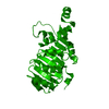
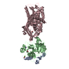
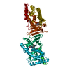
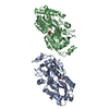
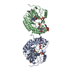
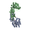
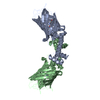
| Title | Structure of crystal form III of TP0453 | ||||||
 Components Components | 30kLP | ||||||
 Keywords Keywords | MEMBRANE PROTEIN / Treponema pallidum / TP0453 / outer membrane protein | ||||||
| Function / homology | : / Outer membrane-associated lipoprotein TP0453 / cell outer membrane / Prokaryotic membrane lipoprotein lipid attachment site profile. / Outer membrane protein TP0453 Function and homology information Function and homology information | ||||||
| Biological species |  Treponema pallidum (bacteria) Treponema pallidum (bacteria) | ||||||
| Method |  X-RAY DIFFRACTION / SYNCHROTRON / MAD / Resolution: 2.2 Å X-RAY DIFFRACTION / SYNCHROTRON / MAD / Resolution: 2.2 Å | ||||||
 Authors Authors | Zhu, G. / Luthra, A. / Desrosiers, D. / Koszelak-Rosenblum, M. / Mulay, V. / Radolf, J.D. / Malkowski, M.G. | ||||||
 Citation Citation | Journal: J.Biol.Chem. / Year: 2011 Title: The Transition from Closed to Open Conformation of Treponema pallidum Outer Membrane-associated Lipoprotein TP0453 Involves Membrane Sensing and Integration by Two Amphipathic Helices. Authors: Luthra, A. / Zhu, G. / Desrosiers, D.C. / Eggers, C.H. / Mulay, V. / Anand, A. / McArthur, F.A. / Romano, F.B. / Caimano, M.J. / Heuck, A.P. / Malkowski, M.G. / Radolf, J.D. | ||||||
| History |
|
- Structure visualization
| Structure viewer | Molecule: MolmilJmol/JSmol |
|---|
- Downloads & links
-Download
| PDBx/mmCIF format | 3k8j.cif.gz | 64.3 KB | Display | PDBx/mmCIF format |
|---|---|---|---|---|
| PDB format | pdb3k8j.ent.gz | 47.4 KB | Display | PDB format |
| PDBx/mmJSON format | 3k8j.json.gz | Tree view | PDBx/mmJSON format | |
| Others |  Other downloads Other downloads |
-Validation report
| Arichive directory | https://data.pdbj.org/pub/pdb/validation_reports/k8/3k8jftp://data.pdbj.org/pub/pdb/validation_reports/k8/3k8j | HTTPS FTP |
|---|
-Related structure data


-Links
 PDBj
PDBj


- Assembly
| Deposited unit | 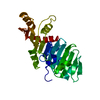
| ||||||||
|---|---|---|---|---|---|---|---|---|---|
| 1 | 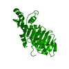
| ||||||||
| Unit cell |
|
-Components
| #1: Protein | Mass: 29147.111 Da / Num. of mol.: 1 / Fragment: residues 27-287 Source method: isolated from a genetically manipulated source Source: (gene. exp.) Treponema pallidum (bacteria) / Gene: 30klp, TP_0453 / Plasmid: pET43.1a / Production host: |
|---|---|
| #2: Water | ChemComp-HOH /  Mass: 18.015 Da / Num. of mol.: 122 / Source method: isolated from a natural source / Formula: H2O Mass: 18.015 Da / Num. of mol.: 122 / Source method: isolated from a natural source / Formula: H2O |
-Experimental details
-Experiment
| Experiment | Method: X-RAY DIFFRACTION / Number of used crystals: 1 |
|---|
- Sample preparation
| Crystal | Density Matthews: 2.65 Å3/Da / Density % sol: 53.67 % |
|---|---|
| Crystal grow | Temperature: 293 K / Method: vapor diffusion, sitting drop / pH: 5.5 Details: 12% PEG 3350, 0.1M MgCl2, 0.1M MES, pH 5.5, 0.5% BOG, VAPOR DIFFUSION, SITTING DROP, temperature 293K |
-Data collection
| Diffraction |
| |||||||||||||||
|---|---|---|---|---|---|---|---|---|---|---|---|---|---|---|---|---|
| Diffraction source |
| |||||||||||||||
| Detector |
| |||||||||||||||
| Radiation | Monochromator: Si 111 / Protocol: SINGLE WAVELENGTH / Monochromatic (M) / Laue (L): M / Scattering type: x-ray | |||||||||||||||
| Radiation wavelength |
| |||||||||||||||
| Reflection | Resolution: 2.2→78.75 Å / Num. all: 16362 / Num. obs: 16362 / % possible obs: 98.6 % / Redundancy: 7 % / Rmerge(I) obs: 0.047 / Net I/σ(I): 43.9 | |||||||||||||||
| Reflection shell | Resolution: 2.2→2.28 Å / Redundancy: 6 % / Rmerge(I) obs: 0.27 / Mean I/σ(I) obs: 6.8 / % possible all: 88.4 |

- Processing
| Software |
| ||||||||||||||||||||||||||||||||||||||||||||||||||||||||||||||||||||||||||||||||||||||||||||||||||||||||||||||||||||||||||||||||||||||||||||||||||||||||||||||||||||||||||
|---|---|---|---|---|---|---|---|---|---|---|---|---|---|---|---|---|---|---|---|---|---|---|---|---|---|---|---|---|---|---|---|---|---|---|---|---|---|---|---|---|---|---|---|---|---|---|---|---|---|---|---|---|---|---|---|---|---|---|---|---|---|---|---|---|---|---|---|---|---|---|---|---|---|---|---|---|---|---|---|---|---|---|---|---|---|---|---|---|---|---|---|---|---|---|---|---|---|---|---|---|---|---|---|---|---|---|---|---|---|---|---|---|---|---|---|---|---|---|---|---|---|---|---|---|---|---|---|---|---|---|---|---|---|---|---|---|---|---|---|---|---|---|---|---|---|---|---|---|---|---|---|---|---|---|---|---|---|---|---|---|---|---|---|---|---|---|---|---|---|---|---|
| Refinement | Method to determine structure: MAD / Resolution: 2.2→78.75 Å / Cor.coef. Fo:Fc: 0.931 / Cor.coef. Fo:Fc free: 0.902 / SU B: 10.001 / SU ML: 0.12 / Cross valid method: THROUGHOUT / ESU R: 0.261 / ESU R Free: 0.216 / Stereochemistry target values: MAXIMUM LIKELIHOOD / Details: HYDROGENS HAVE BEEN ADDED IN THE RIDING POSITIONS
| ||||||||||||||||||||||||||||||||||||||||||||||||||||||||||||||||||||||||||||||||||||||||||||||||||||||||||||||||||||||||||||||||||||||||||||||||||||||||||||||||||||||||||
| Solvent computation | Ion probe radii: 0.8 Å / Shrinkage radii: 0.8 Å / VDW probe radii: 1.4 Å / Solvent model: MASK | ||||||||||||||||||||||||||||||||||||||||||||||||||||||||||||||||||||||||||||||||||||||||||||||||||||||||||||||||||||||||||||||||||||||||||||||||||||||||||||||||||||||||||
| Displacement parameters | Biso mean: 38.527 Å2
| ||||||||||||||||||||||||||||||||||||||||||||||||||||||||||||||||||||||||||||||||||||||||||||||||||||||||||||||||||||||||||||||||||||||||||||||||||||||||||||||||||||||||||
| Refinement step | Cycle: LAST / Resolution: 2.2→78.75 Å
| ||||||||||||||||||||||||||||||||||||||||||||||||||||||||||||||||||||||||||||||||||||||||||||||||||||||||||||||||||||||||||||||||||||||||||||||||||||||||||||||||||||||||||
| Refine LS restraints |
| ||||||||||||||||||||||||||||||||||||||||||||||||||||||||||||||||||||||||||||||||||||||||||||||||||||||||||||||||||||||||||||||||||||||||||||||||||||||||||||||||||||||||||
| LS refinement shell | Resolution: 2.2→2.257 Å / Total num. of bins used: 20
| ||||||||||||||||||||||||||||||||||||||||||||||||||||||||||||||||||||||||||||||||||||||||||||||||||||||||||||||||||||||||||||||||||||||||||||||||||||||||||||||||||||||||||
| Refinement TLS params. | Method: refined / Origin x: 30.7385 Å / Origin y: 25.9564 Å / Origin z: 28.4194 Å
|
