 Movie
Movie Controller

[English] 日本語
 Yorodumi
- PDB-3k7p: Structure of mutant of ribose 5-phosphate isomerase type B from T... -
+ Open data

- Basic information
| Entry | Database: PDB / ID: 3k7p | ||||||
|---|---|---|---|---|---|---|---|
| Title | Structure of mutant of ribose 5-phosphate isomerase type B from Trypanosoma cruzi. | ||||||
 Components Components | Ribose 5-phosphate isomerase | ||||||
 Keywords Keywords | ISOMERASE / pentose phosphate pathway / Type B ribose 5-phosphate isomerase (RpiB) / R5P / phosphate | ||||||
| Function / homology |  Function and homology information Function and homology informationisomerase activity / carbohydrate metabolic process / identical protein binding Similarity search - Function | ||||||
| Biological species |  | ||||||
| Method |  X-RAY DIFFRACTION / SYNCHROTRON / MOLECULAR REPLACEMENT / molecular replacement / Resolution: 1.4 Å X-RAY DIFFRACTION / SYNCHROTRON / MOLECULAR REPLACEMENT / molecular replacement / Resolution: 1.4 Å | ||||||
 Authors Authors | Naworyta, A. / Mowbray, S.L. / Stern, A.L. | ||||||
 Citation Citation | Journal: Febs J. / Year: 2011 Title: Structures of type B ribose 5-phosphate isomerase from Trypanosoma cruzi shed light on the determinants of sugar specificity in the structural family. Authors: Stern, A.L. / Naworyta, A. / Cazzulo, J.J. / Mowbray, S.L. | ||||||
| History |
|
- Structure visualization
| Structure viewer | Molecule: MolmilJmol/JSmol |
|---|
- Downloads & links
-Download
| PDBx/mmCIF format | 3k7p.cif.gz | 75.1 KB | Display | PDBx/mmCIF format |
|---|---|---|---|---|
| PDB format | pdb3k7p.ent.gz | 56 KB | Display | PDB format |
| PDBx/mmJSON format | 3k7p.json.gz | Tree view | PDBx/mmJSON format | |
| Others |  Other downloads Other downloads |
-Validation report
| Arichive directory | https://data.pdbj.org/pub/pdb/validation_reports/k7/3k7pftp://data.pdbj.org/pub/pdb/validation_reports/k7/3k7p | HTTPS FTP |
|---|
-Related structure data
| Related structure data |  3k7oC 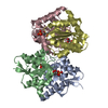 3k7sC 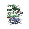 3k8cC 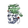 3m1pC 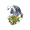 1nn4S C: citing same article ( S: Starting model for refinement |
|---|---|
| Similar structure data |


-Links
 PDBj
PDBj- Assembly
| Deposited unit | 
| ||||||||
|---|---|---|---|---|---|---|---|---|---|
| 1 | 
| ||||||||
| Unit cell |
| ||||||||
| Components on special symmetry positions |
|
-Components
| #1: Protein | Mass: 19527.260 Da / Num. of mol.: 2 / Mutation: C69A Source method: isolated from a genetically manipulated source Source: (gene. exp.)  References: UniProt: A1BTJ7, UniProt: Q4CQE2*PLUS, ribose-5-phosphate isomerase #2: Chemical |   Mass: 94.971 Da / Num. of mol.: 2 / Source method: obtained synthetically / Formula: PO4 Mass: 94.971 Da / Num. of mol.: 2 / Source method: obtained synthetically / Formula: PO4#3: Water | ChemComp-HOH / |  Mass: 18.015 Da / Num. of mol.: 177 / Source method: isolated from a natural source / Formula: H2O Mass: 18.015 Da / Num. of mol.: 177 / Source method: isolated from a natural source / Formula: H2O |
|---|
-Experimental details
-Experiment
| Experiment | Method: X-RAY DIFFRACTION / Number of used crystals: 1 |
|---|
- Sample preparation
| Crystal | Density Matthews: 2.6 Å3/Da / Density % sol: 52.72 % |
|---|---|
| Crystal grow | Temperature: 293 K / Method: vapor diffusion, sitting drop / pH: 8.2 Details: 0.8 M Na/K phosphate, pH 8.2, VAPOR DIFFUSION, SITTING DROP, temperature 293K |
-Data collection
| Diffraction | Mean temperature: 100 K |
|---|---|
| Diffraction source | Source: SYNCHROTRON / Site: ESRF  / Beamline: ID14-1 / Wavelength: 0.934 Å / Beamline: ID14-1 / Wavelength: 0.934 Å |
| Detector | Type: ADSC QUANTUM 210 / Detector: CCD / Date: Oct 27, 2008 |
| Radiation | Protocol: SINGLE WAVELENGTH / Monochromatic (M) / Laue (L): M / Scattering type: x-ray |
| Radiation wavelength | Wavelength: 0.934 Å / Relative weight: 1 |
| Reflection | Resolution: 1.4→30 Å / Num. obs: 79637 / % possible obs: 98.2 % / Observed criterion σ(I): 0 / Redundancy: 5.7 % / Rmerge(I) obs: 0.097 / Rsym value: 0.097 / Net I/σ(I): 11.1 |
| Reflection shell | Resolution: 1.4→1.48 Å / Redundancy: 5.7 % / Rmerge(I) obs: 0.335 / Mean I/σ(I) obs: 3.1 / Num. unique all: 11693 / Rsym value: 0.335 / % possible all: 100 |
-Phasing
| Phasing | Method: molecular replacement | |||||||||
|---|---|---|---|---|---|---|---|---|---|---|
| Phasing MR | Model details: Phaser MODE: MR_AUTO
|
- Processing
| Software |
| ||||||||||||||||||||||||||||||||||||||||||||||||||||||||||||||||||||||||||||||||||||||||||
|---|---|---|---|---|---|---|---|---|---|---|---|---|---|---|---|---|---|---|---|---|---|---|---|---|---|---|---|---|---|---|---|---|---|---|---|---|---|---|---|---|---|---|---|---|---|---|---|---|---|---|---|---|---|---|---|---|---|---|---|---|---|---|---|---|---|---|---|---|---|---|---|---|---|---|---|---|---|---|---|---|---|---|---|---|---|---|---|---|---|---|---|
| Refinement | Method to determine structure: MOLECULAR REPLACEMENT Starting model: pdb entry 1NN4 Resolution: 1.4→30 Å / Cor.coef. Fo:Fc: 0.946 / Cor.coef. Fo:Fc free: 0.939 / WRfactor Rfree: 0.251 / WRfactor Rwork: 0.242 / Occupancy max: 1 / Occupancy min: 0.5 / FOM work R set: 0.856 / SU B: 1.004 / SU ML: 0.041 / SU R Cruickshank DPI: 0.064 / SU Rfree: 0.062 / Cross valid method: THROUGHOUT / σ(F): 0 / ESU R: 0.064 / ESU R Free: 0.062 / Stereochemistry target values: MAXIMUM LIKELIHOOD / Details: HYDROGENS HAVE BEEN ADDED IN THE RIDING POSITIONS
| ||||||||||||||||||||||||||||||||||||||||||||||||||||||||||||||||||||||||||||||||||||||||||
| Solvent computation | Ion probe radii: 0.8 Å / Shrinkage radii: 0.8 Å / VDW probe radii: 1.4 Å / Solvent model: MASK | ||||||||||||||||||||||||||||||||||||||||||||||||||||||||||||||||||||||||||||||||||||||||||
| Displacement parameters | Biso max: 49.84 Å2 / Biso mean: 16.949 Å2 / Biso min: 8.48 Å2
| ||||||||||||||||||||||||||||||||||||||||||||||||||||||||||||||||||||||||||||||||||||||||||
| Refinement step | Cycle: LAST / Resolution: 1.4→30 Å
| ||||||||||||||||||||||||||||||||||||||||||||||||||||||||||||||||||||||||||||||||||||||||||
| Refine LS restraints |
| ||||||||||||||||||||||||||||||||||||||||||||||||||||||||||||||||||||||||||||||||||||||||||
| LS refinement shell | Resolution: 1.4→1.436 Å / Total num. of bins used: 20
|
