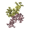
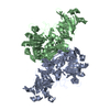
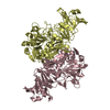
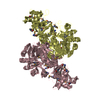
[English] 日本語
Yorodumi
- PDB-3k6m: Dynamic domains of Succinyl-CoA:3-ketoacid-coenzyme A transferase... -

+
Open data


ID or keywords:

Loading...

-
Basic information

Entry
Database: PDB / ID: 3k6m
TitleDynamic domains of Succinyl-CoA:3-ketoacid-coenzyme A transferase from pig heart.
ComponentsSuccinyl-CoA:3-ketoacid-coenzyme A transferase 1, mitochondrial
KeywordsTRANSFERASE / SCOT / CoA Transferase / dynamic domain / glycerol / Mitochondrion / Transit peptide
Function / homology
Function and homology information


Utilization of Ketone Bodies / cellular ketone body metabolic process / 3-oxoacid CoA-transferase / succinyl-CoA:3-oxo-acid CoA-transferase activity / ketone body catabolic process / Mitochondrial protein degradation / protein homodimerization activity / mitochondrion
Similarity search - Function
3-oxoacid CoA-transferase, subunit A / Coenzyme A transferase active site / 3-oxoacid CoA-transferase, subunit B / 3-oxoacid CoA-transferase / Coenzyme A transferases signature 2. / Coenzyme A transferase binding site / Coenzyme A transferases signature 1. / Glutaconate Coenzyme A-transferase / Glutaconate Coenzyme A-transferase / Coenzyme A transferase family I ...3-oxoacid CoA-transferase, subunit A / Coenzyme A transferase active site / 3-oxoacid CoA-transferase, subunit B / 3-oxoacid CoA-transferase / Coenzyme A transferases signature 2. / Coenzyme A transferase binding site / Coenzyme A transferases signature 1. / Glutaconate Coenzyme A-transferase / Glutaconate Coenzyme A-transferase / Coenzyme A transferase family I / Coenzyme A transferase / Coenzyme A transferase / NagB/RpiA transferase-like / 3-Layer(aba) Sandwich / Alpha Beta
Similarity search - Domain/homology
Succinyl-CoA:3-ketoacid coenzyme A transferase 1, mitochondrial
Similarity search - Component
Biological speciesSus scrofa (pig)
MethodX-RAY DIFFRACTION / SYNCHROTRON / MOLECULAR REPLACEMENT / Resolution: 1.5 Å
AuthorsCoker, S. / Lloyd, A. / Mitchell, E. / Lewis, G.R. / Shoolingin-Jordan, P. / Coker, A.R.
CitationJournal: Acta Crystallogr.,Sect.D / Year: 2010
Title: The high-resolution structure of pig heart succinyl-CoA:3-oxoacid coenzyme A transferase.
Authors: Coker, S.F. / Lloyd, A.J. / Mitchell, E. / Lewis, G.R. / Coker, A.R. / Shoolingin-Jordan, P.M.
History
DepositionOct 9, 2009Deposition site: RCSB / Processing site: RCSB
Revision 1.0Jul 7, 2010Provider: repository / Type: Initial release
Revision 1.1Jul 13, 2011Group: Advisory / Refinement description / Version format compliance
Revision 1.2Mar 21, 2012Group: Database references
Revision 1.3Sep 6, 2023Group: Data collection / Database references ...Data collection / Database references / Derived calculations / Refinement description
Category: chem_comp_atom / chem_comp_bond ...chem_comp_atom / chem_comp_bond / database_2 / pdbx_initial_refinement_model / struct_site
Item: _database_2.pdbx_DOI / _database_2.pdbx_database_accession ..._database_2.pdbx_DOI / _database_2.pdbx_database_accession / _struct_site.pdbx_auth_asym_id / _struct_site.pdbx_auth_comp_id / _struct_site.pdbx_auth_seq_id

-
Structure visualization

Structure viewerMolecule:
MolmilJmol/JSmol

Downloads & links

-
Assembly

Deposited unit
A: Succinyl-CoA:3-ketoacid-coenzyme A transferase 1, mitochondrial
D: Succinyl-CoA:3-ketoacid-coenzyme A transferase 1, mitochondrial
C: Succinyl-CoA:3-ketoacid-coenzyme A transferase 1, mitochondrial
B: Succinyl-CoA:3-ketoacid-coenzyme A transferase 1, mitochondrial
hetero molecules


Theoretical massNumber of molelcules
Total (without water)209,4318
Polymers209,1764
Non-polymers2554
Water28,4821581
1
A: Succinyl-CoA:3-ketoacid-coenzyme A transferase 1, mitochondrial
B: Succinyl-CoA:3-ketoacid-coenzyme A transferase 1, mitochondrial
hetero molecules


Theoretical massNumber of molelcules
Total (without water)104,6233
Polymers104,5882
Non-polymers351
Water362
TypeNameSymmetry operationNumber
identity operation1_555x,y,z1
Buried area4640 Å2
ΔGint-14 kcal/mol
Surface area34540 Å2
MethodPISA
2
D: Succinyl-CoA:3-ketoacid-coenzyme A transferase 1, mitochondrial
C: Succinyl-CoA:3-ketoacid-coenzyme A transferase 1, mitochondrial
hetero molecules


Theoretical massNumber of molelcules
Total (without water)104,8085
Polymers104,5882
Non-polymers2203
Water362
TypeNameSymmetry operationNumber
identity operation1_555x,y,z1
Buried area4610 Å2
ΔGint-14 kcal/mol
Surface area33880 Å2
MethodPISA
Unit cell
Length a, b, c (Å)73.715, 133.566, 102.230
Angle α, β, γ (deg.)90.00, 104.98, 90.00
Int Tables number4
Space group name H-MP1211

-
Components

#1: Protein
Succinyl-CoA:3-ketoacid-coenzyme A transferase 1, mitochondrial / 3-oxoacid-CoA transferase 1 / Somatic-type succinyl CoA:3-oxoacid CoA-transferase / Scot-S


Mass: 52293.957 Da / Num. of mol.: 4 / Fragment: residues 40-520 / Source method: isolated from a natural source / Details: heart / Source: (natural) Sus scrofa (pig) / References: UniProt: Q29551, 3-oxoacid CoA-transferase
#2: Chemical ChemComp-CL / CHLORIDE ION / Chloride


Mass: 35.453 Da / Num. of mol.: 2 / Source method: obtained synthetically / Formula: Cl
#3: Chemical ChemComp-GOL / GLYCEROL / GLYCERIN / PROPANE-1,2,3-TRIOL / Glycerol


Mass: 92.094 Da / Num. of mol.: 2 / Source method: obtained synthetically / Formula: C3H8O3
#4: Water ChemComp-HOH / water / Water


Mass: 18.015 Da / Num. of mol.: 1581 / Source method: isolated from a natural source / Formula: H2O

-
Experimental details

-
Experiment

ExperimentMethod: X-RAY DIFFRACTION / Number of used crystals: 1

-
Sample preparation

CrystalDensity Matthews: 2.32 Å3/Da / Density % sol: 47.08 %
Crystal growTemperature: 277 K / Method: vapor diffusion, hanging drop / pH: 8
Details: 4 ul Well Solution: 18-22% PEG 3350 or 4000, 75 mM Tris/HCl pH 8. 4 ul Protein solution: 10-15 mg/ml protein, 20 mM MOPS pH 7.2, 1 mM EDTA, 1 mM DTT, 0.2 mM PMSF, 10% glycerol, VAPOR ...Details: 4 ul Well Solution: 18-22% PEG 3350 or 4000, 75 mM Tris/HCl pH 8. 4 ul Protein solution: 10-15 mg/ml protein, 20 mM MOPS pH 7.2, 1 mM EDTA, 1 mM DTT, 0.2 mM PMSF, 10% glycerol, VAPOR DIFFUSION, HANGING DROP, temperature 277K

-
Data collection

DiffractionMean temperature: 100 K
Diffraction sourceSource: SYNCHROTRON / Site: ESRF / Beamline: ID14-3 / Wavelength: 0.948 Å
DetectorType: MAR CCD 130 mm / Detector: CCD / Date: Jun 25, 1998
RadiationProtocol: SINGLE WAVELENGTH / Monochromatic (M) / Laue (L): M / Scattering type: x-ray
Radiation wavelengthWavelength: 0.948 Å / Relative weight: 1
ReflectionResolution: 1.5→20 Å / Num. all: 297163 / Num. obs: 297163 / % possible obs: 97.7 % / Observed criterion σ(F): 0 / Observed criterion σ(I): 0 / Redundancy: 3.3 % / Biso Wilson estimate: 17.16 Å2 / Rmerge(I) obs: 0.042 / Rsym value: 0.042 / Net I/σ(I): 12
Reflection shellResolution: 1.5→1.54 Å / Redundancy: 2.2 % / Rmerge(I) obs: 0.296 / Mean I/σ(I) obs: 2.3 / Num. unique all: 18372 / Rsym value: 0.296 / % possible all: 82

-
Processing

Software
NameVersionClassification
AMoREphasing
REFMAC5.2.0019refinement
MOSFLMdata reduction
SCALAdata scaling
RefinementMethod to determine structure: MOLECULAR REPLACEMENT
Starting model: PDB ENTRY 1O9L
Resolution: 1.5→20 Å / Cor.coef. Fo:Fc: 0.971 / Cor.coef. Fo:Fc free: 0.963 / SU B: 2.485 / SU ML: 0.047 / TLS residual ADP flag: LIKELY RESIDUAL / Isotropic thermal model: ISOTROPIC / Cross valid method: THROUGHOUT / σ(F): 0 / ESU R: 0.069 / ESU R Free: 0.068 / Stereochemistry target values: MAXIMUM LIKELIHOOD / Details: HYDROGENS HAVE BEEN ADDED IN THE RIDING POSITIONS
RfactorNum. reflection% reflectionSelection details
Rfree0.18538 14899 5 %RANDOM
Rwork0.16502 ---
all0.1657 296782 --
obs0.1657 296782 97.6 %-
Solvent computationIon probe radii: 0.8 Å / Shrinkage radii: 0.8 Å / VDW probe radii: 1.4 Å / Solvent model: BABINET MODEL WITH MASK
Displacement parametersBiso mean: 19.924 Å2
Baniso -1Baniso -2Baniso -3
1-0.92 Å20 Å20.15 Å2
2---0.76 Å20 Å2
3----0.08 Å2
Refine analyzeLuzzati coordinate error obs: 0.238 Å
Refinement stepCycle: LAST / Resolution: 1.5→20 Å
ProteinNucleic acidLigandSolventTotal
Num. atoms14137 0 14 1581 15732
Refine LS restraints
Refine-IDTypeDev idealDev ideal targetNumber
X-RAY DIFFRACTIONr_bond_refined_d0.0160.02214752
X-RAY DIFFRACTIONr_bond_other_d00.0210089
X-RAY DIFFRACTIONr_angle_refined_deg1.5281.97719997
X-RAY DIFFRACTIONr_angle_other_deg4.206324849
X-RAY DIFFRACTIONr_dihedral_angle_1_deg6.20651964
X-RAY DIFFRACTIONr_dihedral_angle_2_deg38.6824.817602
X-RAY DIFFRACTIONr_dihedral_angle_3_deg12.414152698
X-RAY DIFFRACTIONr_dihedral_angle_4_deg13.9021577
X-RAY DIFFRACTIONr_chiral_restr0.0970.22296
X-RAY DIFFRACTIONr_gen_planes_refined0.0070.0216464
X-RAY DIFFRACTIONr_gen_planes_other0.0090.022801
X-RAY DIFFRACTIONr_nbd_refined0.220.22550
X-RAY DIFFRACTIONr_nbd_other0.2420.210168
X-RAY DIFFRACTIONr_nbtor_refined0.1770.27197
X-RAY DIFFRACTIONr_nbtor_other0.1110.27353
X-RAY DIFFRACTIONr_xyhbond_nbd_refined0.170.2829
X-RAY DIFFRACTIONr_symmetry_vdw_refined0.1750.285
X-RAY DIFFRACTIONr_symmetry_vdw_other0.3040.2306
X-RAY DIFFRACTIONr_symmetry_hbond_refined0.1520.2365
X-RAY DIFFRACTIONr_mcbond_it1.1141.512235
X-RAY DIFFRACTIONr_mcbond_other0.231.53891
X-RAY DIFFRACTIONr_mcangle_it1.208215095
X-RAY DIFFRACTIONr_scbond_it2.31536179
X-RAY DIFFRACTIONr_scangle_it3.2314.54848
LS refinement shellResolution: 1.5→1.539 Å / Total num. of bins used: 20
RfactorNum. reflection% reflection
Rfree0.242 910 -
Rwork0.225 17341 -
obs-17341 81.77 %
Refinement TLS params.

Method: refined / Refine-ID: X-RAY DIFFRACTION

IDL112)L122)L132)L222)L232)L332)S11 (Å °)S12 (Å °)S13 (Å °)S21 (Å °)S22 (Å °)S23 (Å °)S31 (Å °)S32 (Å °)S33 (Å °)T112)T122)T132)T222)T232)T332)Origin x (Å)Origin y (Å)Origin z (Å)
10.6349-0.0283-0.06020.7053-0.08130.86360.02030.01990.07840.0379-0.0092-0.0459-0.1748-0.0432-0.0112-0.02550.0490.0022-0.12010.0103-0.158418.113766.918756.5878
23.23940.6332-0.01770.51450.22671.4694-0.0447-0.11350.0340.02090.0855-0.16110.03690.2971-0.0408-0.00890.0442-0.0354-0.0074-0.0115-0.030648.893868.018962.2421
30.5362-0.0896-0.04330.782-0.15410.8143-0.00570.042-0.084-0.03450.0036-0.04270.0794-0.04420.0021-0.07920.02680.0037-0.1162-0.0084-0.141220.541935.671455.7122
42.42830.4078-0.92041.3971-0.50682.6438-0.06240.2949-0.1003-0.10930.06130.25380.2113-0.41060.0011-0.07710.0033-0.00020.0185-0.0276-0.0659-9.923834.699865.1475
50.64920.03980.05280.97290.14640.6986-0.017-0.01370.0781-0.02040.02550.0229-0.08060.0279-0.0085-0.2093-0.0051-0.0055-0.1904-0.0066-0.14225.120273.85734.5221
62.60830.492-0.31811.60750.42051.73810.00810.07190.0512-0.08180.0325-0.245-0.12620.2223-0.0405-0.1899-0.0310.0037-0.0935-0.0077-0.056734.493477.38361.0751
70.7962-0.19310.03430.93710.1740.8349-0.0194-0.0654-0.11270.06160.02760.03010.13380.0247-0.0082-0.19260.0042-0.0015-0.18520.0024-0.13427.343442.81245.1388
82.5942-0.13240.10981.574-1.00893.3006-0.0713-0.396-0.20320.17130.19760.4220.2148-0.6889-0.1263-0.0929-0.05190.10490.09230.06340.0659-19.63842.520421.3651
Refinement TLS group
IDRefine-IDRefine TLS-IDAuth asym-IDAuth seq-ID
1X-RAY DIFFRACTION1A1 - 246
2X-RAY DIFFRACTION1A300 - 358
3X-RAY DIFFRACTION1A375 - 394
4X-RAY DIFFRACTION2A262 - 299
5X-RAY DIFFRACTION2A359 - 374
6X-RAY DIFFRACTION2A395 - 480
7X-RAY DIFFRACTION3B1 - 246
8X-RAY DIFFRACTION3B300 - 358
9X-RAY DIFFRACTION3B375 - 394
10X-RAY DIFFRACTION4B261 - 299
11X-RAY DIFFRACTION4B359 - 374
12X-RAY DIFFRACTION4B395 - 480
13X-RAY DIFFRACTION5C1 - 246
14X-RAY DIFFRACTION5C300 - 358
15X-RAY DIFFRACTION5C375 - 394
16X-RAY DIFFRACTION6C261 - 299
17X-RAY DIFFRACTION6C359 - 374
18X-RAY DIFFRACTION6C395 - 480
19X-RAY DIFFRACTION7D1 - 246
20X-RAY DIFFRACTION7D300 - 358
21X-RAY DIFFRACTION7D375 - 394
22X-RAY DIFFRACTION8D262 - 299
23X-RAY DIFFRACTION8D359 - 374
24X-RAY DIFFRACTION8D395 - 480

+
About Yorodumi

-
News

-
Feb 9, 2022. New format data for meta-information of EMDB entries

New format data for meta-information of EMDB entries

  • Version 3 of the EMDB header file is now the official format.
  • The previous official version 1.9 will be removed from the archive.

Related info.:EMDB header

External links:wwPDB to switch to version 3 of the EMDB data model

-
Aug 12, 2020. Covid-19 info

Covid-19 info

URL: https://pdbj.org/emnavi/covid19.php

New page: Covid-19 featured information page in EM Navigator.

Related info.:Covid-19 info / Mar 5, 2020. Novel coronavirus structure data

+
Mar 5, 2020. Novel coronavirus structure data

Novel coronavirus structure data

Related info.:Yorodumi Speices / Aug 12, 2020. Covid-19 info

External links:COVID-19 featured content - PDBj / Molecule of the Month (242):Coronavirus Proteases

+
Jan 31, 2019. EMDB accession codes are about to change! (news from PDBe EMDB page)

EMDB accession codes are about to change! (news from PDBe EMDB page)

  • The allocation of 4 digits for EMDB accession codes will soon come to an end. Whilst these codes will remain in use, new EMDB accession codes will include an additional digit and will expand incrementally as the available range of codes is exhausted. The current 4-digit format prefixed with “EMD-” (i.e. EMD-XXXX) will advance to a 5-digit format (i.e. EMD-XXXXX), and so on. It is currently estimated that the 4-digit codes will be depleted around Spring 2019, at which point the 5-digit format will come into force.
  • The EM Navigator/Yorodumi systems omit the EMD- prefix.

Related info.:Q: What is EMD? / ID/Accession-code notation in Yorodumi/EM Navigator

External links:EMDB Accession Codes are Changing Soon! / Contact to PDBj

+
Jul 12, 2017. Major update of PDB

Major update of PDB

  • wwPDB released updated PDB data conforming to the new PDBx/mmCIF dictionary.
  • This is a major update changing the version number from 4 to 5, and with Remediation, in which all the entries are updated.
  • In this update, many items about electron microscopy experimental information are reorganized (e.g. em_software).
  • Now, EM Navigator and Yorodumi are based on the updated data.

External links:wwPDB Remediation / Enriched Model Files Conforming to OneDep Data Standards Now Available in the PDB FTP Archive

-
Yorodumi

Thousand views of thousand structures

  • Yorodumi is a browser for structure data from EMDB, PDB, SASBDB, etc.
  • This page is also the successor to EM Navigator detail page, and also detail information page/front-end page for Omokage search.
  • The word "yorodu" (or yorozu) is an old Japanese word meaning "ten thousand". "mi" (miru) is to see.

Related info.:EMDB / PDB / SASBDB / Comparison of 3 databanks / Yorodumi Search / Aug 31, 2016. New EM Navigator & Yorodumi / Yorodumi Papers / Jmol/JSmol / Function and homology information / Changes in new EM Navigator and Yorodumi

Read more