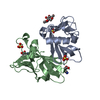
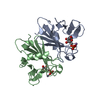
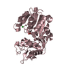
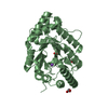
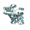
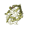
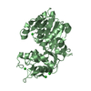
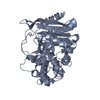
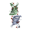
Crystal structure of sCD-MPR mutant E19Q/K137M pH 7.0
Components
Cation-dependent mannose-6-phosphate receptor
Keywords
protein transport / sugar binding protein / transport / lysosome / mannose / receptor / sugar binding / Glycoprotein / Membrane / Phosphoprotein / Transmembrane
Function / homology
Function and homology information
Retrograde transport at the Trans-Golgi-Network / Glycosphingolipid catabolism / secretion of lysosomal enzymes / Lysosome Vesicle Biogenesis / retromer complex binding / Cargo recognition for clathrin-mediated endocytosis / Clathrin-mediated endocytosis / protein targeting to lysosome / lysosomal transport / trans-Golgi network ...Retrograde transport at the Trans-Golgi-Network / Glycosphingolipid catabolism / secretion of lysosomal enzymes / Lysosome Vesicle Biogenesis / retromer complex binding / Cargo recognition for clathrin-mediated endocytosis / Clathrin-mediated endocytosis / protein targeting to lysosome / lysosomal transport / trans-Golgi network / late endosome / endosome / protein domain specific binding / lysosomal membrane / perinuclear region of cytoplasm / Golgi apparatus Similarity search - Function
In the structure databanks used in Yorodumi, some data are registered as the other names, "COVID-19 virus" and "2019-nCoV". Here are the details of the virus and the list of structure data.
Jan 31, 2019. EMDB accession codes are about to change! (news from PDBe EMDB page)
EMDB accession codes are about to change! (news from PDBe EMDB page)
The allocation of 4 digits for EMDB accession codes will soon come to an end. Whilst these codes will remain in use, new EMDB accession codes will include an additional digit and will expand incrementally as the available range of codes is exhausted. The current 4-digit format prefixed with “EMD-” (i.e. EMD-XXXX) will advance to a 5-digit format (i.e. EMD-XXXXX), and so on. It is currently estimated that the 4-digit codes will be depleted around Spring 2019, at which point the 5-digit format will come into force.
The EM Navigator/Yorodumi systems omit the EMD- prefix.
Related info.:Q: What is EMD? / ID/Accession-code notation in Yorodumi/EM Navigator
Yorodumi is a browser for structure data from EMDB, PDB, SASBDB, etc.
This page is also the successor to EM Navigator detail page, and also detail information page/front-end page for Omokage search.
The word "yorodu" (or yorozu) is an old Japanese word meaning "ten thousand". "mi" (miru) is to see.
Related info.:EMDB / PDB / SASBDB / Comparison of 3 databanks / Yorodumi Search / Aug 31, 2016. New EM Navigator & Yorodumi / Yorodumi Papers / Jmol/JSmol / Function and homology information / Changes in new EM Navigator and Yorodumi
 Movie
Movie Controller


 Open data
 Basic information
 Components
 Keywords
 Function and homology information
 X-RAY DIFFRACTION /
  Authors
 Citation
 Structure visualization
 Downloads & links
 Other downloads
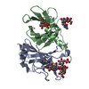


 PDBj
PDBj Assembly
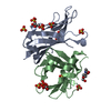
 Pichia Pastoris (fungus) / Strain (production host): x-33 / References: UniProt: P11456
 Type: D-saccharide, beta linking / Mass: 221.208 Da / Num. of mol.: 2 / Source method: obtained synthetically / Formula: C8H15NO6
 Mass: 172.074 Da / Num. of mol.: 2 / Source method: obtained synthetically / Formula: C3H9O6P
 Mass: 96.063 Da / Num. of mol.: 8 / Source method: obtained synthetically / Formula: SO4
 Mass: 18.015 Da / Num. of mol.: 180 / Source method: isolated from a natural source / Formula: H2O
 Sample preparation
 Processing