 Movie
Movie Controller


+ Open data

- Basic information
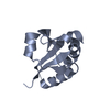
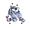
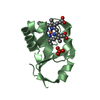
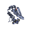
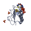
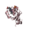
| Entry | Database: PDB / ID: 3jtn | ||||||
|---|---|---|---|---|---|---|---|
| Title | Crystal Structure of the c-terminal domain of YpbH | ||||||
 Components Components | Adapter protein mecA 2 | ||||||
 Keywords Keywords | PROTEIN BINDING / YpbH / adaptor protein / Competence / Sporulation | ||||||
| Function / homology |  Function and homology information Function and homology informationnegative regulation of establishment of competence for transformation / negative regulation of sporulation resulting in formation of a cellular spore / establishment of competence for transformation / sporulation resulting in formation of a cellular spore / protein-macromolecule adaptor activity Similarity search - Function | ||||||
| Biological species |  | ||||||
| Method |  X-RAY DIFFRACTION / PHASER / Resolution: 2.09 Å X-RAY DIFFRACTION / PHASER / Resolution: 2.09 Å | ||||||
 Authors Authors | Wang, F. / Mei, Z. / Qi, Y. / Yan, C. / Wang, J. / Shi, Y. | ||||||
 Citation Citation | Journal: To be Published Title: Crystal Structure of the MecA degradation tag Authors: Wang, F. / Mei, Z. / Qi, Y. / Yan, C. / Xiang, S. / Zhou, Z. / Hu, Q. / Wang, J. / Shi, Y. | ||||||
| History |
|
- Structure visualization
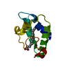
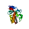
| Structure viewer | Molecule: MolmilJmol/JSmol |
|---|
- Downloads & links
-Download
| PDBx/mmCIF format | 3jtn.cif.gz | 82.8 KB | Display | PDBx/mmCIF format |
|---|---|---|---|---|
| PDB format | pdb3jtn.ent.gz | 64.8 KB | Display | PDB format |
| PDBx/mmJSON format | 3jtn.json.gz | Tree view | PDBx/mmJSON format | |
| Others |  Other downloads Other downloads |
-Validation report
| Arichive directory | https://data.pdbj.org/pub/pdb/validation_reports/jt/3jtnftp://data.pdbj.org/pub/pdb/validation_reports/jt/3jtn | HTTPS FTP |
|---|
-Related structure data
| Related structure data | |
|---|---|
| Similar structure data |


-Links
 PDBj
PDBj- Assembly
| Deposited unit | 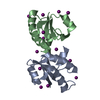
| ||||||||
|---|---|---|---|---|---|---|---|---|---|
| 1 | 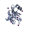
| ||||||||
| 2 | 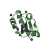
| ||||||||
| Unit cell |
|
-Components
| #1: Protein | Mass: 10182.252 Da / Num. of mol.: 2 / Fragment: c-terminal domain, UNP residues 104-194 Source method: isolated from a genetically manipulated source Source: (gene. exp.) #2: Chemical | ChemComp-IOD /   Mass: 126.904 Da / Num. of mol.: 14 / Source method: obtained synthetically / Formula: I Mass: 126.904 Da / Num. of mol.: 14 / Source method: obtained synthetically / Formula: I#3: Water | ChemComp-HOH / |  Mass: 18.015 Da / Num. of mol.: 60 / Source method: isolated from a natural source / Formula: H2O Mass: 18.015 Da / Num. of mol.: 60 / Source method: isolated from a natural source / Formula: H2O |
|---|
-Experimental details
-Experiment
| Experiment | Method: X-RAY DIFFRACTION / Number of used crystals: 1 |
|---|
- Sample preparation
| Crystal | Density Matthews: 1.99 Å3/Da / Density % sol: 38.29 % |
|---|---|
| Crystal grow | Temperature: 291 K / Method: vapor diffusion, hanging drop / pH: 6.5 Details: 20% PEG 3350,0.3M calcium chloride, 0.1M Bis-Tris pH6.5, VAPOR DIFFUSION, HANGING DROP, temperature 291K |
-Data collection
| Diffraction | Mean temperature: 100 K |
|---|---|
| Diffraction source | Source: ROTATING ANODE / Type: RIGAKU MICROMAX-007 HF / Wavelength: 1.5418 Å |
| Detector | Type: RIGAKU RAXIS HTC / Detector: IMAGE PLATE / Date: Jul 22, 2009 / Details: mirrors |
| Radiation | Protocol: SINGLE WAVELENGTH / Monochromatic (M) / Laue (L): M / Scattering type: x-ray |
| Radiation wavelength | Wavelength: 1.5418 Å / Relative weight: 1 |
| Reflection | Resolution: 2.09→34.363 Å / Num. all: 17571 / Num. obs: 17571 / % possible obs: 94.1 % / Observed criterion σ(F): 0 / Observed criterion σ(I): 0 / Redundancy: 2.9 % / Biso Wilson estimate: 30.99 Å2 / Rmerge(I) obs: 0.079 / Net I/σ(I): 9.09 |
| Reflection shell | Resolution: 2.09→2.21 Å / Redundancy: 2.7 % / Rmerge(I) obs: 0.732 / Mean I/σ(I) obs: 2.6 / Num. unique all: 17571 / % possible all: 88 |
- Processing
| Software |
| |||||||||||||||||||||||||||||||||||||||||||||||||||||||||||||||||||||||||||
|---|---|---|---|---|---|---|---|---|---|---|---|---|---|---|---|---|---|---|---|---|---|---|---|---|---|---|---|---|---|---|---|---|---|---|---|---|---|---|---|---|---|---|---|---|---|---|---|---|---|---|---|---|---|---|---|---|---|---|---|---|---|---|---|---|---|---|---|---|---|---|---|---|---|---|---|---|
| Refinement | Method to determine structure: PHASER / Resolution: 2.09→34.363 Å / Occupancy max: 1 / Occupancy min: 0.21 / FOM work R set: 0.698 / SU ML: 0.35 / σ(F): 0.03 / Phase error: 36.01 / Stereochemistry target values: ML Details: The file contains friedel pairs in the _refln.pdbx_F_plus and _refln.pdbx_F_minus columns.
| |||||||||||||||||||||||||||||||||||||||||||||||||||||||||||||||||||||||||||
| Solvent computation | Shrinkage radii: 0.9 Å / VDW probe radii: 1.11 Å / Solvent model: FLAT BULK SOLVENT MODEL / Bsol: 37.434 Å2 / ksol: 0.388 e/Å3 | |||||||||||||||||||||||||||||||||||||||||||||||||||||||||||||||||||||||||||
| Displacement parameters | Biso max: 226.24 Å2 / Biso mean: 43.401 Å2 / Biso min: 15.09 Å2
| |||||||||||||||||||||||||||||||||||||||||||||||||||||||||||||||||||||||||||
| Refinement step | Cycle: LAST / Resolution: 2.09→34.363 Å
| |||||||||||||||||||||||||||||||||||||||||||||||||||||||||||||||||||||||||||
| Refine LS restraints |
| |||||||||||||||||||||||||||||||||||||||||||||||||||||||||||||||||||||||||||
| LS refinement shell | Refine-ID: X-RAY DIFFRACTION / Total num. of bins used: 6
| |||||||||||||||||||||||||||||||||||||||||||||||||||||||||||||||||||||||||||
| Refinement TLS params. | Method: refined / Refine-ID: X-RAY DIFFRACTION
| |||||||||||||||||||||||||||||||||||||||||||||||||||||||||||||||||||||||||||
| Refinement TLS group |
|
