 Movie
Movie Controller

[English] 日本語
 Yorodumi
- PDB-3jdw: CRYSTAL STRUCTURE AND MECHANISM OF L-ARGININE: GLYCINE AMIDINOTRA... -
+ Open data

- Basic information
| Entry | Database: PDB / ID: 3jdw | ||||||
|---|---|---|---|---|---|---|---|
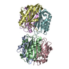
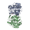
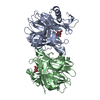
| Title | CRYSTAL STRUCTURE AND MECHANISM OF L-ARGININE: GLYCINE AMIDINOTRANSFERASE: A MITOCHONDRIAL ENZYME INVOLVED IN CREATINE BIOSYNTHESIS | ||||||
 Components Components | L-ARGININE\:GLYCINE AMIDINOTRANSFERASE | ||||||
 Keywords Keywords | TRANSFERASE / CREATINE BIOSYNTHESIS / CATALYTIC TRIAD / REACTION MECHANISM / NOVEL FOLD / FIVEFOLD PSEUDOSYMMETRY | ||||||
| Function / homology |  Function and homology information Function and homology informationglycine amidinotransferase / glycine amidinotransferase activity / amidinotransferase activity / creatine biosynthetic process / creatine metabolic process / muscle atrophy / Creatine metabolism / mitochondrial intermembrane space / positive regulation of cold-induced thermogenesis / learning or memory ...glycine amidinotransferase / glycine amidinotransferase activity / amidinotransferase activity / creatine biosynthetic process / creatine metabolic process / muscle atrophy / Creatine metabolism / mitochondrial intermembrane space / positive regulation of cold-induced thermogenesis / learning or memory / mitochondrial inner membrane / mitochondrion / extracellular exosome Similarity search - Function | ||||||
| Biological species |  Homo sapiens (human) Homo sapiens (human) | ||||||
| Method |  X-RAY DIFFRACTION / ISOMORPHOUS WITH PDB ENTRY 1JDW / Resolution: 2.4 Å X-RAY DIFFRACTION / ISOMORPHOUS WITH PDB ENTRY 1JDW / Resolution: 2.4 Å | ||||||
 Authors Authors | Humm, A. / Fritsche, E. / Steinbacher, S. / Huber, R. | ||||||
 Citation Citation | Journal: EMBO J. / Year: 1997 Title: Crystal structure and mechanism of human L-arginine:glycine amidinotransferase: a mitochondrial enzyme involved in creatine biosynthesis. Authors: Humm, A. / Fritsche, E. / Steinbacher, S. / Huber, R. #1: Journal: Biol.Chem.Hoppe-Seyler / Year: 1997Title: Structure and Reaction Mechanism of L-Arginine:Glycine Amidinotransferase Authors: Humm, A. / Fritsche, E. / Steinbacher, S. #2: Journal: Eur.J.Biochem. / Year: 1997Title: Substrate Binding and Catalysis by L-Arginine:Glycine Amidinotransferase--A Mutagenesis and Crystallographic Study Authors: Fritsche, E. / Humm, A. / Huber, R. #3: Journal: Biochem.J. / Year: 1997Title: Recombinant Expression and Isolation of Human L-Arginine:Glycine Amidinotransferase and Identification of its Active-Site Cysteine Residue Authors: Humm, A. / Fritsche, E. / Mann, K. / Gohl, M. / Huber, R. #4: Journal: J.Mol.Biol. / Year: 1997Title: Bioincorporation of Telluromethionine Into Proteins: A Promising New Approach for X-Ray Structure Analysis of Proteins Authors: Budisa, N. / Karnbrock, W. / Steinbacher, S. / Humm, A. / Prade, L. / Neuefeind, T. / Moroder, L. / Huber, R. | ||||||
| History |
|
- Structure visualization
| Structure viewer | Molecule: MolmilJmol/JSmol |
|---|
- Downloads & links
-Download
| PDBx/mmCIF format | 3jdw.cif.gz | 111.1 KB | Display | PDBx/mmCIF format |
|---|---|---|---|---|
| PDB format | pdb3jdw.ent.gz | 86.6 KB | Display | PDB format |
| PDBx/mmJSON format | 3jdw.json.gz | Tree view | PDBx/mmJSON format | |
| Others |  Other downloads Other downloads |
-Validation report
| Arichive directory | https://data.pdbj.org/pub/pdb/validation_reports/jd/3jdwftp://data.pdbj.org/pub/pdb/validation_reports/jd/3jdw | HTTPS FTP |
|---|
-Related structure data
| Related structure data |  1jdwSC  2jdwC  4jdwC S: Starting model for refinement C: citing same article ( |
|---|---|
| Similar structure data |


-Links
 PDBj
PDBj- Assembly
| Deposited unit | 
| ||||||||
|---|---|---|---|---|---|---|---|---|---|
| 1 | 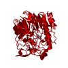
| ||||||||
| Unit cell |
|
-Components
| #1: Protein | Mass: 48521.367 Da / Num. of mol.: 1 / Fragment: RESIDUES 64 - 423 Source method: isolated from a genetically manipulated source Source: (gene. exp.) Homo sapiens (human)Description: WITHOUT SIGNAL SEQUENCE (1-37) BUT WITH N-TERMINAL ATTACHED 6-HISTIDINE-TAG (14 RESIDUES) Cell line: BL21 Cellular location: INTERMEMBRANE SPACE OF MITOCHONDRIA AND CYTOPLASM Gene: AT38H / Organ: KIDNEY / Organelle: MITOCHONDRIA / Plasmid: PRSETAT38H / Cellular location (production host): CYTOSOLIC / Gene (production host): AT38H / Production host:  |
|---|---|
| #2: Chemical | ChemComp-ORN /   Type: L-peptide linking / Mass: 132.161 Da / Num. of mol.: 1 / Source method: obtained synthetically / Formula: C5H12N2O2 Type: L-peptide linking / Mass: 132.161 Da / Num. of mol.: 1 / Source method: obtained synthetically / Formula: C5H12N2O2 |
| #3: Water | ChemComp-HOH /  Mass: 18.015 Da / Num. of mol.: 193 / Source method: isolated from a natural source / Formula: H2O Mass: 18.015 Da / Num. of mol.: 193 / Source method: isolated from a natural source / Formula: H2O |
-Experimental details
-Experiment
| Experiment | Method: X-RAY DIFFRACTION / Number of used crystals: 1 |
|---|
- Sample preparation
| Crystal | Density Matthews: 3.8 Å3/Da / Density % sol: 65 % | |||||||||||||||||||||||||
|---|---|---|---|---|---|---|---|---|---|---|---|---|---|---|---|---|---|---|---|---|---|---|---|---|---|---|
| Crystal grow | pH: 7 Details: 1 PART OF AT38H (13-16 MG/ML) 2 PARTS OF PRECIPITANT (3% PEG 6000, 40 MM HEPES, 1MM GLUTATHIONE, PH7.0), SEVERAL DAYS AT ROOM TEMPERATURE, MACRO SEEDING. Temp details: room temp | |||||||||||||||||||||||||
| Crystal grow | *PLUS Temperature: 22 ℃ / Method: vapor diffusion | |||||||||||||||||||||||||
| Components of the solutions | *PLUS
|
-Data collection
| Diffraction | Mean temperature: 290 K |
|---|---|
| Diffraction source | Source: ROTATING ANODE / Type: RIGAKU RUH2R / Wavelength: 1.5418 |
| Detector | Type: MARRESEARCH / Detector: IMAGE PLATE / Date: Nov 11, 1995 |
| Radiation | Monochromator: GRAPHITE(002) / Monochromatic (M) / Laue (L): M / Scattering type: x-ray |
| Radiation wavelength | Wavelength: 1.5418 Å / Relative weight: 1 |
| Reflection | Resolution: 2.4→8 Å / Num. obs: 25838 / % possible obs: 93.1 % / Observed criterion σ(I): 0 / Redundancy: 3.78 % / Rmerge(I) obs: 0.074 |
| Reflection shell | Resolution: 2.4→2.43 Å / Redundancy: 1.7 % / Rmerge(I) obs: 0.287 / % possible all: 51.9 |
| Reflection shell | *PLUS % possible obs: 51.9 % |
- Processing
| Software |
| ||||||||||||||||||||||||||||||||||||||||||||||||||||||||||||
|---|---|---|---|---|---|---|---|---|---|---|---|---|---|---|---|---|---|---|---|---|---|---|---|---|---|---|---|---|---|---|---|---|---|---|---|---|---|---|---|---|---|---|---|---|---|---|---|---|---|---|---|---|---|---|---|---|---|---|---|---|---|
| Refinement | Method to determine structure: ISOMORPHOUS WITH PDB ENTRY 1JDW Starting model: PDB ENTRY 1JDW Resolution: 2.4→8 Å / Cross valid method: FREE R / σ(F): 0 Details: RESIDUES 38 - 63 ARE NOT VISIBLE IN THE ELECTRON DENSITY
| ||||||||||||||||||||||||||||||||||||||||||||||||||||||||||||
| Displacement parameters | Biso mean: 27.61 Å2 | ||||||||||||||||||||||||||||||||||||||||||||||||||||||||||||
| Refinement step | Cycle: LAST / Resolution: 2.4→8 Å
| ||||||||||||||||||||||||||||||||||||||||||||||||||||||||||||
| Refine LS restraints |
| ||||||||||||||||||||||||||||||||||||||||||||||||||||||||||||
| LS refinement shell | Resolution: 2.4→2.43 Å / Total num. of bins used: 30
| ||||||||||||||||||||||||||||||||||||||||||||||||||||||||||||
| Software | *PLUS Name: X-PLOR / Version: 3.1 / Classification: refinement | ||||||||||||||||||||||||||||||||||||||||||||||||||||||||||||
| Refine LS restraints | *PLUS
|
