 Movie
Movie Controller


+ Open data

- Basic information
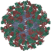
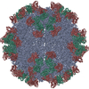
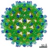
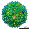
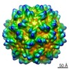
| Entry | Database: PDB / ID: 3jcx | ||||||
|---|---|---|---|---|---|---|---|
| Title | Canine Parvovirus complexed with Fab E | ||||||
 Components Components |
| ||||||
 Keywords Keywords | VIRUS/IMMUNE SYSTEM /  parvovirus / antibody / VIRUS-IMMUNE SYSTEM complex parvovirus / antibody / VIRUS-IMMUNE SYSTEM complex | ||||||
| Function / homology |  Function and homology information Function and homology informationpermeabilization of host organelle membrane involved in viral entry into host cell / symbiont entry into host cell via permeabilization of inner membrane / T=1 icosahedral viral capsid / clathrin-dependent endocytosis of virus by host cell / virion attachment to host cell / structural molecule activity Similarity search - Function | ||||||
| Biological species |  Canine parvovirus Canine parvovirus Rattus norvegicus (Norway rat) Rattus norvegicus (Norway rat) | ||||||
| Method | ELECTRON MICROSCOPY / single particle reconstruction / cryo EM / Resolution: 4.1 Å | ||||||
 Authors Authors | Organtini, L.J. / Iketani, S. / Huang, K. / Ashley, R.E. / Makhov, A.M. / Conway, J.F. / Parrish, C.R. / Hafenstein, S. | ||||||
 Citation Citation | Journal: J Virol / Year: 2016 Title: Near-Atomic Resolution Structure of a Highly Neutralizing Fab Bound to Canine Parvovirus. Authors: Lindsey J Organtini / Hyunwook Lee / Sho Iketani / Kai Huang / Robert E Ashley / Alexander M Makhov / James F Conway / Colin R Parrish / Susan Hafenstein /  Abstract: Canine parvovirus (CPV) is a highly contagious pathogen that causes severe disease in dogs and wildlife. Previously, a panel of neutralizing monoclonal antibodies (MAb) raised against CPV was ...Canine parvovirus (CPV) is a highly contagious pathogen that causes severe disease in dogs and wildlife. Previously, a panel of neutralizing monoclonal antibodies (MAb) raised against CPV was characterized. An antibody fragment (Fab) of MAb E was found to neutralize the virus at low molar ratios. Using recent advances in cryo-electron microscopy (cryo-EM), we determined the structure of CPV in complex with Fab E to 4.1 Å resolution, which allowed de novo building of the Fab structure. The footprint identified was significantly different from the footprint obtained previously from models fitted into lower-resolution maps. Using single-chain variable fragments, we tested antibody residues that control capsid binding. The near-atomic structure also revealed that Fab binding had caused capsid destabilization in regions containing key residues conferring receptor binding and tropism, which suggests a mechanism for efficient virus neutralization by antibody. Furthermore, a general technical approach to solving the structures of small molecules is demonstrated, as binding the Fab to the capsid allowed us to determine the 50-kDa Fab structure by cryo-EM. IMPORTANCE: Using cryo-electron microscopy and new direct electron detector technology, we have solved the 4 Å resolution structure of a Fab molecule bound to a picornavirus capsid. The Fab induced ...IMPORTANCE: Using cryo-electron microscopy and new direct electron detector technology, we have solved the 4 Å resolution structure of a Fab molecule bound to a picornavirus capsid. The Fab induced conformational changes in regions of the virus capsid that control receptor binding. The antibody footprint is markedly different from the previous one identified by using a 12 Å structure. This work emphasizes the need for a high-resolution structure to guide mutational analysis and cautions against relying on older low-resolution structures even though they were interpreted with the best methodology available at the time. | ||||||
| History |
|
- Structure visualization
| Movie |
Movie viewer |
|---|---|
| Structure viewer | Molecule: MolmilJmol/JSmol |
- Downloads & links
-Download
| PDBx/mmCIF format | 3jcx.cif.gz | 148.8 KB | Display | PDBx/mmCIF format |
|---|---|---|---|---|
| PDB format | pdb3jcx.ent.gz | 118.7 KB | Display | PDB format |
| PDBx/mmJSON format | 3jcx.json.gz | Tree view | PDBx/mmJSON format | |
| Others |  Other downloads Other downloads |
-Validation report
| Arichive directory | https://data.pdbj.org/pub/pdb/validation_reports/jc/3jcxftp://data.pdbj.org/pub/pdb/validation_reports/jc/3jcx | HTTPS FTP |
|---|
-Related structure data
| Related structure data |  6629MC M: map data used to model this data C: citing same article ( |
|---|---|
| Similar structure data |
-Links
 PDBj
PDBj


- Assembly
| Deposited unit | 
|
|---|---|
| 1 | x 60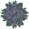
|
| 2 |
|
| 3 | x 5
|
| 4 | x 6
|
| 5 | 
|
| Symmetry | Point symmetry: (Schoenflies symbol: I (icosahedral)) |
-Components
| #1: Protein | / VP2 Mass: 64783.629 Da / Num. of mol.: 1 / Source method: isolated from a natural source / Source: (natural) Canine parvovirus / References: UniProt: B2ZG07 |
|---|---|
| #2: Antibody | Mass: 12739.242 Da / Num. of mol.: 1 / Source method: isolated from a natural source / Source: (natural) Rattus norvegicus (Norway rat) / Strain: B5A8 |
| #3: Antibody | Mass: 11745.118 Da / Num. of mol.: 1 / Source method: isolated from a natural source / Source: (natural) Rattus norvegicus (Norway rat) / Strain: B5A8 |
-Experimental details
-Experiment
| Experiment | Method: ELECTRON MICROSCOPY |
|---|---|
| EM experiment | Aggregation state: PARTICLE / 3D reconstruction method: single particle reconstruction |
- Sample preparation
| Component |
| |||||||||||||||||||||||||
|---|---|---|---|---|---|---|---|---|---|---|---|---|---|---|---|---|---|---|---|---|---|---|---|---|---|---|
| Details of virus | Empty: YES / Enveloped: NO / Host category: CANINES / Isolate: SPECIES / Type: VIRION | |||||||||||||||||||||||||
| Natural host | Organism: Canis lupus | |||||||||||||||||||||||||
| Buffer solution | Name: PBS / pH: 7.4 / Details: PBS | |||||||||||||||||||||||||
| Specimen | Conc.: 1 mg/ml / Embedding applied: NO / Shadowing applied: NO / Staining applied: NO / Vitrification applied: YES | |||||||||||||||||||||||||
| Specimen support | Details: Quantifoil | |||||||||||||||||||||||||
| Vitrification | Instrument: FEI VITROBOT MARK II / Cryogen name: ETHANE / Details: Plunged into liquid ethane (FEI VITROBOT MARK II). |
- Electron microscopy imaging
| Experimental equipment |  Model: Tecnai Polara / Image courtesy: FEI Company |
|---|---|
| Microscopy | Model: FEI POLARA 300 / Date: Dec 20, 2014 |
| Electron gun | Electron source: FIELD EMISSION GUN / Accelerating voltage: 300 kV / Illumination mode: FLOOD BEAM |
| Electron lens | Mode: BRIGHT FIELDBright-field microscopy / Nominal defocus max: 4000 nm / Nominal defocus min: 1500 nm |
| Specimen holder | Specimen holder model: SIDE ENTRY, EUCENTRIC |
| Image recording | Electron dose: 30 e/Å2 / Film or detector model: FEI FALCON II (4k x 4k) |
| Image scans | Num. digital images: 1424 |
- Processing
| EM software | Name: RELION / Category: 3D reconstruction | ||||||||||||
|---|---|---|---|---|---|---|---|---|---|---|---|---|---|
| Symmetry | Point symmetry: I (icosahedral) | ||||||||||||
| 3D reconstruction | Method: Single ParticleSingle particle analysis / Resolution: 4.1 Å / Resolution method: FSC 0.143 CUT-OFF / Num. of particles: 47563 / Nominal pixel size: 1.37 Å / Actual pixel size: 1.37 Å / Symmetry type: POINT | ||||||||||||
| Refinement step | Cycle: LAST
|
