 Movie
Movie Controller

[English] 日本語
 Yorodumi
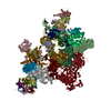
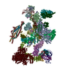
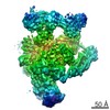
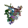
- PDB-3jcr: 3D structure determination of the human*U4/U6.U5* tri-snRNP complex -
+ Open data

- Basic information
| Entry | Database: PDB / ID: 3jcr | ||||||
|---|---|---|---|---|---|---|---|
| Title | 3D structure determination of the human*U4/U6.U5* tri-snRNP complex | ||||||
 Components Components |
| ||||||
 Keywords Keywords | SPLICING / snRNP / spliceosome / human | ||||||
| Function / homology |  Function and homology information Function and homology informationLsm2-8 complex / U6 snRNA 3'-end binding / spliceosomal snRNP complex / ribonucleoprotein complex localization / U4atac snRNP / RNA localization / U4/U6 snRNP / U4atac snRNA binding / mRNA decay by 5' to 3' exoribonuclease / Lsm1-7-Pat1 complex ...Lsm2-8 complex / U6 snRNA 3'-end binding / spliceosomal snRNP complex / ribonucleoprotein complex localization / U4atac snRNP / RNA localization / U4/U6 snRNP / U4atac snRNA binding / mRNA decay by 5' to 3' exoribonuclease / Lsm1-7-Pat1 complex / R-loop processing / box C/D sno(s)RNA binding / U6 snRNP / PH domain binding / U2 snRNP binding / U7 snRNA binding / rRNA 2'-O-methylation / histone pre-mRNA DCP binding / dense fibrillar component / U7 snRNP / cis assembly of pre-catalytic spliceosome / histone pre-mRNA 3'end processing complex / deadenylation-dependent decapping of nuclear-transcribed mRNA / nuclear histone mRNA catabolic process / SLBP independent Processing of Histone Pre-mRNAs / SLBP Dependent Processing of Replication-Dependent Histone Pre-mRNAs / box C/D methylation guide snoRNP complex / 7-methylguanosine cap hypermethylation / U12-type spliceosomal complex / U1 snRNP binding / pICln-Sm protein complex / U2-type catalytic step 1 spliceosome / methylosome / RNA splicing, via transesterification reactions / sno(s)RNA-containing ribonucleoprotein complex / small nuclear ribonucleoprotein complex / SMN-Sm protein complex / protein methylation / spliceosomal tri-snRNP complex / P granule / commitment complex / U4 snRNP / snRNP binding / U2-type precatalytic spliceosome / U2-type prespliceosome assembly / U2-type catalytic step 2 spliceosome / U2-type spliceosomal complex / telomerase holoenzyme complex / U4 snRNA binding / box C/D snoRNP assembly / telomerase RNA binding / spliceosome conformational change to release U4 (or U4atac) and U1 (or U11) / U1 snRNP / U2 snRNP / U3 snoRNA binding / RNA Polymerase II Transcription Termination / P-body assembly / U2-type prespliceosome / tRNA processing / K63-linked polyubiquitin modification-dependent protein binding / mRNA modification / precatalytic spliceosome / rRNA modification in the nucleus and cytosol / mRNA catabolic process / mRNA Splicing - Minor Pathway / MLL1 complex / spliceosomal complex assembly / negative regulation of mRNA splicing, via spliceosome / spliceosomal tri-snRNP complex assembly / U5 snRNP / U5 snRNA binding / pre-mRNA intronic binding / U2 snRNA binding / U6 snRNA binding / protein deubiquitination / ribonucleoprotein complex binding / Cajal body / U1 snRNA binding / RNA processing / U4/U6 x U5 tri-snRNP complex / spliceosomal snRNP assembly / Major pathway of rRNA processing in the nucleolus and cytosol / catalytic step 2 spliceosome / mRNA Polyadenylation / mRNA Splicing - Major Pathway / RNA splicing / helicase activity / spliceosomal complex / response to cocaine / maturation of SSU-rRNA / cellular response to xenobiotic stimulus / small-subunit processome / cellular response to tumor necrosis factor / mRNA splicing, via spliceosome / P-body / Dengue Virus-Host Interactions / small GTPase binding / osteoblast differentiation / neuron differentiation / mRNA processing Similarity search - Function | ||||||
| Biological species |  Homo sapiens (human) Homo sapiens (human) | ||||||
| Method | ELECTRON MICROSCOPY / single particle reconstruction / cryo EM / Resolution: 7 Å | ||||||
 Authors Authors | Agafonov, D.E. / Kastner, B. / Dybkov, O. / Hofele, R.V. / Liu, W.T. / Urlaub, H. / Luhrmann, R. / Stark, H. | ||||||
 Citation Citation | Journal: Science / Year: 2016 Title: Molecular architecture of the human U4/U6.U5 tri-snRNP. Authors: Dmitry E Agafonov / Berthold Kastner / Olexandr Dybkov / Romina V Hofele / Wen-Ti Liu / Henning Urlaub / Reinhard Lührmann / Holger Stark /  Abstract: The U4/U6.U5 triple small nuclear ribonucleoprotein (tri-snRNP) is a major spliceosome building block. We obtained a three-dimensional structure of the 1.8-megadalton human tri-snRNP at a resolution ...The U4/U6.U5 triple small nuclear ribonucleoprotein (tri-snRNP) is a major spliceosome building block. We obtained a three-dimensional structure of the 1.8-megadalton human tri-snRNP at a resolution of 7 angstroms using single-particle cryo-electron microscopy (cryo-EM). We fit all known high-resolution structures of tri-snRNP components into the EM density map and validated them by protein cross-linking. Our model reveals how the spatial organization of Brr2 RNA helicase prevents premature U4/U6 RNA unwinding in isolated human tri-snRNPs and how the ubiquitin C-terminal hydrolase-like protein Sad1 likely tethers the helicase Brr2 to its preactivation position. Comparison of our model with cryo-EM three-dimensional structures of the Saccharomyces cerevisiae tri-snRNP and Schizosaccharomyces pombe spliceosome indicates that Brr2 undergoes a marked conformational change during spliceosome activation, and that the scaffolding protein Prp8 is also rearranged to accommodate the spliceosome's catalytic RNA network. | ||||||
| History |
|
- Structure visualization
| Movie |
Movie viewer |
|---|---|
| Structure viewer | Molecule: MolmilJmol/JSmol |
- Downloads & links
-Download
| PDBx/mmCIF format | 3jcr.cif.gz | 392.6 KB | Display | PDBx/mmCIF format |
|---|---|---|---|---|
| PDB format | pdb3jcr.ent.gz | 185.8 KB | Display | PDB format |
| PDBx/mmJSON format | 3jcr.json.gz | Tree view | PDBx/mmJSON format | |
| Others |  Other downloads Other downloads |
-Validation report
| Arichive directory | https://data.pdbj.org/pub/pdb/validation_reports/jc/3jcrftp://data.pdbj.org/pub/pdb/validation_reports/jc/3jcr | HTTPS FTP |
|---|
-Related structure data
| Related structure data | 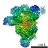 6581MC M: map data used to model this data C: citing same article ( |
|---|---|
| Similar structure data | |
| EM raw data | EMPIAR-10056 (Title: Structure of the human U4/U6.U5 tri-snRNP / Data size: 300.9 / Data #1: TBD [micrographs - single frame]) |


-Links
 PDBj
PDBj


- Assembly
| Deposited unit | 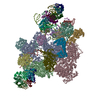
|
|---|---|
| 1 |
|
-Components
+Protein , 26 types, 33 molecules GDCEAFBOoPpQqRrSsTtUu8654327KL...
-RNA chain , 3 types, 3 molecules MNH
| #27: RNA chain | Mass: 46536.492 Da / Num. of mol.: 1 / Source method: isolated from a natural source / Source: (natural) Homo sapiens (human) |
|---|---|
| #28: RNA chain | Mass: 34098.270 Da / Num. of mol.: 1 / Source method: isolated from a natural source / Source: (natural) Homo sapiens (human) |
| #29: RNA chain | Mass: 36891.684 Da / Num. of mol.: 1 / Source method: isolated from a natural source / Source: (natural) Homo sapiens (human) |
-Experimental details
-Experiment
| Experiment | Method: ELECTRON MICROSCOPY |
|---|---|
| EM experiment | Aggregation state: PARTICLE / 3D reconstruction method: single particle reconstruction |
- Sample preparation
| Component |
| |||||||||||||||
|---|---|---|---|---|---|---|---|---|---|---|---|---|---|---|---|---|
| Molecular weight | Value: 1.8 MDa / Experimental value: YES | |||||||||||||||
| Buffer solution | Name: 20 mM HEPES, pH 7.9, 100 mM KCl, 5 mM MgCl2, 0.1 mM EDTA pH: 7.9 Details: 20 mM HEPES, pH 7.9, 100 mM KCl, 5 mM MgCl2, 0.1 mM EDTA | |||||||||||||||
| Specimen | Conc.: 0.1 mg/ml / Embedding applied: NO / Shadowing applied: NO / Staining applied: NO / Vitrification applied: YES | |||||||||||||||
| Specimen support | Details: 200 mesh copper grid with carbon support film | |||||||||||||||
| Vitrification | Instrument: FEI VITROBOT MARK IV / Cryogen name: ETHANE / Humidity: 95 % / Details: Plunged into liquid ethane (FEI VITROBOT MARK IV). |
- Electron microscopy imaging
| Experimental equipment |  Model: Titan Krios / Image courtesy: FEI Company |
|---|---|
| Microscopy | Model: FEI TITAN KRIOS / Date: Sep 10, 2015 |
| Electron gun | Electron source:  FIELD EMISSION GUN / Accelerating voltage: 300 kV / Illumination mode: FLOOD BEAM FIELD EMISSION GUN / Accelerating voltage: 300 kV / Illumination mode: FLOOD BEAM |
| Electron lens | Mode: BRIGHT FIELD / Calibrated magnification: 74000 X / Nominal defocus max: 5350 nm / Nominal defocus min: 1000 nm / Cs: 0.0001 mm |
| Specimen holder | Specimen holder model: FEI TITAN KRIOS AUTOGRID HOLDER |
| Image recording | Electron dose: 45 e/Å2 / Film or detector model: FEI FALCON II (4k x 4k) |
| Image scans | Num. digital images: 4688 |
- Processing
| EM software | Name: RELION / Category: 3D reconstruction | ||||||||||||
|---|---|---|---|---|---|---|---|---|---|---|---|---|---|
| Symmetry | Point symmetry: C1 (asymmetric) | ||||||||||||
| 3D reconstruction | Resolution: 7 Å / Resolution method: FSC 0.143 CUT-OFF / Num. of particles: 141109 / Nominal pixel size: 2 Å / Actual pixel size: 2 Å / Details: (Single particle--Applied symmetry: C1) / Symmetry type: POINT | ||||||||||||
| Refinement step | Cycle: LAST
|
