+ データを開く
- 基本情報
基本情報
| 登録情報 | データベース: PDB / ID: 3jba | ||||||
|---|---|---|---|---|---|---|---|
| タイトル | The U4 antibody epitope on human papillomavirus 16 identified by cryo-EM | ||||||
 要素 要素 |
| ||||||
 キーワード キーワード | VIRUS/IMMUNE SYSTEM / HPV16 / antibody / U4 / neutralization / Fab / VIRUS-IMMUNE SYSTEM complex | ||||||
| 機能・相同性 |  機能・相同性情報 機能・相同性情報T=7 icosahedral viral capsid / endocytosis involved in viral entry into host cell / virion attachment to host cell / host cell nucleus / structural molecule activity 類似検索 - 分子機能 | ||||||
| 生物種 |  Human papillomavirus type 16 (パピローマウイルス) Human papillomavirus type 16 (パピローマウイルス) | ||||||
| 手法 | 電子顕微鏡法 / 単粒子再構成法 / クライオ電子顕微鏡法 / 解像度: 12 Å | ||||||
 データ登録者 データ登録者 | Guan, J. / Bywaters, S.M. / Brendle, S.A. / Lee, H. / Ashley, R.E. / Christensen, N.D. / Hafenstein, S. | ||||||
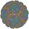
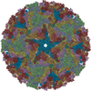
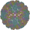
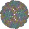
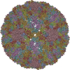
 引用 引用 |  ジャーナル: J Virol / 年: 2015 ジャーナル: J Virol / 年: 2015タイトル: The U4 Antibody Epitope on Human Papillomavirus 16 Identified by Cryo-electron Microscopy. 著者: Jian Guan / Stephanie M Bywaters / Sarah A Brendle / Hyunwook Lee / Robert E Ashley / Neil D Christensen / Susan Hafenstein /  要旨: The human papillomavirus (HPV) major structural protein L1 composes capsomers that are linked together through interactions mediated by the L1 C terminus to constitute a T=7 icosahedral capsid. H16. ...The human papillomavirus (HPV) major structural protein L1 composes capsomers that are linked together through interactions mediated by the L1 C terminus to constitute a T=7 icosahedral capsid. H16.U4 is a type-specific monoclonal antibody recognizing a conformation-dependent neutralizing epitope of HPV thought to include the L1 protein C terminus. The structure of human papillomavirus 16 (HPV16) complexed with H16.U4 fragments of antibody (Fab) was solved by cryo-electron microscopy (cryo-EM) image reconstruction. Atomic structures of virus and Fab were fitted into the corresponding cryo-EM densities to identify the antigenic epitope. The antibody footprint mapped predominately to the L1 C-terminal arm with an additional contact point on the side of the capsomer. This footprint describes an epitope that is presented capsid-wide. However, although the H16.U4 epitope suggests the presence of 360 potential binding sites exposed in the capsid valley between each capsomer, H16.U4 Fab bound only to epitopes located around the icosahedral five-fold vertex of the capsid. Thus, the binding characteristics of H16.U4 defined in this study showed a distinctive selectivity for local conformation-dependent interactions with specific L1 invading arms between five-fold related capsomers. IMPORTANCE: Human papillomavirus 16 (HPV16) is the most prevalent oncogenic genotype in HPV-associated anogenital and oral cancers. Here we use cryo-EM reconstruction techniques to solve the ...IMPORTANCE: Human papillomavirus 16 (HPV16) is the most prevalent oncogenic genotype in HPV-associated anogenital and oral cancers. Here we use cryo-EM reconstruction techniques to solve the structures of the HPV16 capsid complexes using H16.U4 fragment of antibody (Fab). Different from most other antibodies directed against surface loops, H16.U4 monoclonal antibody is unique in targeting the C-terminal arm of the L1 protein. This monoclonal antibody (MAb) is used throughout the HPV research community in HPV serological and vaccine development and to define mechanisms of HPV uptake. The unique binding mode of H16.U4 defined here shows important conformation-dependent interactions within the HPV16 capsid. By targeting an important structural and conformational epitope, H16.U4 may identify subtle conformational changes in different maturation stages of the HPV capsid and provide a key probe to analyze the mechanisms of HPV uptake during the early stages of virus infection. Our analyses precisely define important conformational epitopes on HPV16 capsids that are key targets for successful HPV prophylactic vaccines. | ||||||
| 履歴 |
|
- 構造の表示
構造の表示
| ムービー |
 ムービービューア ムービービューア |
|---|---|
| 構造ビューア | 分子:  Molmil Molmil Jmol/JSmol Jmol/JSmol |
- ダウンロードとリンク
- ダウンロード
| PDBx/mmCIF形式 |  3jba.cif.gz 3jba.cif.gz | 541.7 KB | 表示 |  PDBx/mmCIF形式 PDBx/mmCIF形式 |
|---|---|---|---|---|
| PDB形式 |  pdb3jba.ent.gz pdb3jba.ent.gz | 359.9 KB | 表示 |  PDB形式 PDB形式 |
| PDBx/mmJSON形式 |  3jba.json.gz 3jba.json.gz | ツリー表示 |  PDBx/mmJSON形式 PDBx/mmJSON形式 | |
| その他 |  その他のダウンロード その他のダウンロード |
-検証レポート
| 文書・要旨 |  3jba_validation.pdf.gz 3jba_validation.pdf.gz | 813 KB | 表示 |  wwPDB検証レポート wwPDB検証レポート |
|---|---|---|---|---|
| 文書・詳細版 |  3jba_full_validation.pdf.gz 3jba_full_validation.pdf.gz | 839 KB | 表示 | |
| XML形式データ |  3jba_validation.xml.gz 3jba_validation.xml.gz | 67.6 KB | 表示 | |
| CIF形式データ |  3jba_validation.cif.gz 3jba_validation.cif.gz | 115.8 KB | 表示 | |
| アーカイブディレクトリ |  https://data.pdbj.org/pub/pdb/validation_reports/jb/3jba https://data.pdbj.org/pub/pdb/validation_reports/jb/3jba ftp://data.pdbj.org/pub/pdb/validation_reports/jb/3jba ftp://data.pdbj.org/pub/pdb/validation_reports/jb/3jba | HTTPS FTP |
-関連構造データ
- リンク
リンク
- 集合体
集合体
| 登録構造単位 | 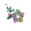
|
|---|---|
| 1 | x 60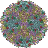
|
| 2 |
|
| 3 | x 5
|
| 4 | x 6
|
| 5 | 
|
| 対称性 | 点対称性: (シェーンフリース記号: I (正20面体型対称)) |
- 要素
要素
| #1: 抗体 | 分子量: 12699.254 Da / 分子数: 1 / 断片: Fab variable domain / 由来タイプ: 天然 / 由来: (天然)  | ||
|---|---|---|---|
| #2: 抗体 | 分子量: 12711.025 Da / 分子数: 1 / 断片: Fab variable domain / 由来タイプ: 天然 / 由来: (天然)  | ||
| #3: タンパク質 | 分子量: 53445.617 Da / 分子数: 6 / 由来タイプ: 組換発現 由来: (組換発現)  Human papillomavirus type 16 (パピローマウイルス) Human papillomavirus type 16 (パピローマウイルス)遺伝子: L1 / プラスミド: SV40 / 細胞株 (発現宿主): HEK293TT / 発現宿主:  Homo sapiens (ヒト) / 参照: UniProt: C9E771, UniProt: P03101*PLUS Homo sapiens (ヒト) / 参照: UniProt: C9E771, UniProt: P03101*PLUSHas protein modification | Y | |
-実験情報
-実験
| 実験 | 手法: 電子顕微鏡法 |
|---|---|
| EM実験 | 試料の集合状態: PARTICLE / 3次元再構成法: 単粒子再構成法 |
- 試料調製
試料調製
| 構成要素 |
| ||||||||||||||||
|---|---|---|---|---|---|---|---|---|---|---|---|---|---|---|---|---|---|
| 分子量 | 値: 32.8 MDa / 実験値: NO | ||||||||||||||||
| ウイルスについての詳細 | 中空か: NO / エンベロープを持つか: NO / ホストのカテゴリ: VERTEBRATES / 単離: SEROTYPE / タイプ: VIRUS-LIKE PARTICLE | ||||||||||||||||
| 天然宿主 | 生物種: Homo sapiens | ||||||||||||||||
| 緩衝液 | 名称: 1 M NaCl, 200 mM Tris / pH: 7.4 / 詳細: 1 M NaCl, 200 mM Tris | ||||||||||||||||
| 試料 | 濃度: 1.2 mg/ml / 包埋: NO / シャドウイング: NO / 染色: NO / 凍結: YES | ||||||||||||||||
| 試料支持 | 詳細: glow-discharged holey carbon support grid | ||||||||||||||||
| 急速凍結 | 装置: GATAN CRYOPLUNGE 3 / 凍結剤: ETHANE / Temp: 102 K / 湿度: 90 % / 詳細: Plunged into liquid ethane (GATAN CRYOPLUNGE 3). |
- 電子顕微鏡撮影
| 顕微鏡 | モデル: JEOL 2100 / 日付: 2014年7月22日 |
|---|---|
| 電子銃 | 電子線源: LAB6 / 加速電圧: 200 kV / 照射モード: SPOT SCAN |
| 電子レンズ | モード: BRIGHT FIELD / 倍率(公称値): 50000 X / 最大 デフォーカス(公称値): 5430 nm / 最小 デフォーカス(公称値): 1600 nm / Cs: 2 mm |
| 試料ホルダ | 試料ホルダーモデル: GATAN LIQUID NITROGEN / 温度: 95 K |
| 撮影 | 電子線照射量: 15 e/Å2 フィルム・検出器のモデル: GATAN ULTRASCAN 4000 (4k x 4k) |
| 画像スキャン | デジタル画像の数: 151 |
| 放射 | プロトコル: SINGLE WAVELENGTH / 単色(M)・ラウエ(L): M / 散乱光タイプ: x-ray |
| 放射波長 | 相対比: 1 |
- 解析
解析
| EMソフトウェア |
| ||||||||||||||||||||||||
|---|---|---|---|---|---|---|---|---|---|---|---|---|---|---|---|---|---|---|---|---|---|---|---|---|---|
| CTF補正 | 詳細: each particle | ||||||||||||||||||||||||
| 対称性 | 点対称性: I (正20面体型対称) | ||||||||||||||||||||||||
| 3次元再構成 | 手法: Cross-common lines / 解像度: 12 Å / 解像度の算出法: FSC 0.5 CUT-OFF / 粒子像の数: 5806 / ピクセルサイズ(公称値): 2.33 Å / ピクセルサイズ(実測値): 2.33 Å 詳細: (Single particle details: The particles were selected using semi-automatic program e2boxer.py.) (Single particle--Applied symmetry: I) 対称性のタイプ: POINT | ||||||||||||||||||||||||
| 原子モデル構築 | プロトコル: RIGID BODY FIT / 空間: REAL / 詳細: REFINEMENT PROTOCOL--rigid body | ||||||||||||||||||||||||
| 原子モデル構築 | PDB-ID: 3J6R PDB chain-ID: F / Accession code: 3J6R / Source name: PDB / タイプ: experimental model | ||||||||||||||||||||||||
| 精密化ステップ | サイクル: LAST
|
 ムービー
ムービー コントローラー


 UCSF Chimera


 PDBj
PDBj


