+ データを開く
- 基本情報
基本情報
| 登録情報 | データベース: PDB / ID: 3jao | ||||||
|---|---|---|---|---|---|---|---|
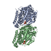
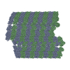
| タイトル | Ciliary microtubule doublet | ||||||
 要素 要素 |
| ||||||
 キーワード キーワード | STRUCTURAL PROTEIN / tubulin / microtubule doublet / cilia | ||||||
| 機能・相同性 |  機能・相同性情報 機能・相同性情報microtubule-based process / structural constituent of cytoskeleton / microtubule / hydrolase activity / GTPase activity / GTP binding 類似検索 - 分子機能 | ||||||
| 生物種 |  | ||||||
| 手法 | 電子顕微鏡法 / 単粒子再構成法 / クライオ電子顕微鏡法 / 解像度: 23 Å | ||||||
 データ登録者 データ登録者 | Maheshwari, A. / Obbineni, J.M. / Bui, K.H. / Shibata, K. / Toyoshima, Y.Y. / Ishikawa, T. | ||||||
 引用 引用 |  ジャーナル: Structure / 年: 2015 ジャーナル: Structure / 年: 2015タイトル: α- and β-Tubulin Lattice of the Axonemal Microtubule Doublet and Binding Proteins Revealed by Single Particle Cryo-Electron Microscopy and Tomography. 著者: Aditi Maheshwari / Jagan Mohan Obbineni / Khanh Huy Bui / Keitaro Shibata / Yoko Y Toyoshima / Takashi Ishikawa /   要旨: Microtubule doublet (MTD) is the main skeleton of cilia/flagella. Many proteins, such as dyneins and radial spokes, bind to MTD, and generate or regulate force. While the structure of the ...Microtubule doublet (MTD) is the main skeleton of cilia/flagella. Many proteins, such as dyneins and radial spokes, bind to MTD, and generate or regulate force. While the structure of the reconstituted microtubule has been solved at atomic resolution, nature of the axonemal MTD is still unclear. There are a few hypotheses of the lattice arrangement of its α- and β-tubulins, but it has not been described how dyneins and radial spokes bind to MTD. In this study, we analyzed the three-dimensional structure of Tetrahymena MTD at ∼19 Å resolution by single particle cryo-electron microscopy. To identify α- and β-tubulins, we combined image analysis of MTD with specific kinesin decoration. This work reveals that α- and β-tubulins form a B-lattice arrangement in the entire MTD with a seam at the outer junction. We revealed the unique way in which inner arm dyneins, radial spokes, and proteins inside MTD bind and bridge protofilaments. | ||||||
| 履歴 |
|
- 構造の表示
構造の表示
| ムービー |
 ムービービューア ムービービューア |
|---|---|
| 構造ビューア | 分子:  Molmil Molmil Jmol/JSmol Jmol/JSmol |
- ダウンロードとリンク
- ダウンロード
| PDBx/mmCIF形式 |  3jao.cif.gz 3jao.cif.gz | 170.3 KB | 表示 |  PDBx/mmCIF形式 PDBx/mmCIF形式 |
|---|---|---|---|---|
| PDB形式 |  pdb3jao.ent.gz pdb3jao.ent.gz | 128.4 KB | 表示 |  PDB形式 PDB形式 |
| PDBx/mmJSON形式 |  3jao.json.gz 3jao.json.gz | ツリー表示 |  PDBx/mmJSON形式 PDBx/mmJSON形式 | |
| その他 |  その他のダウンロード その他のダウンロード |
-検証レポート
| 文書・要旨 |  3jao_validation.pdf.gz 3jao_validation.pdf.gz | 922.5 KB | 表示 |  wwPDB検証レポート wwPDB検証レポート |
|---|---|---|---|---|
| 文書・詳細版 |  3jao_full_validation.pdf.gz 3jao_full_validation.pdf.gz | 941.4 KB | 表示 | |
| XML形式データ |  3jao_validation.xml.gz 3jao_validation.xml.gz | 32.1 KB | 表示 | |
| CIF形式データ |  3jao_validation.cif.gz 3jao_validation.cif.gz | 47.4 KB | 表示 | |
| アーカイブディレクトリ |  https://data.pdbj.org/pub/pdb/validation_reports/ja/3jao https://data.pdbj.org/pub/pdb/validation_reports/ja/3jao ftp://data.pdbj.org/pub/pdb/validation_reports/ja/3jao ftp://data.pdbj.org/pub/pdb/validation_reports/ja/3jao | HTTPS FTP |
-関連構造データ
- リンク
リンク
- 集合体
集合体
| 登録構造単位 | 
|
|---|---|
| 1 | x 115
|
| 2 |
|
| 3 | 
|
- 要素
要素
-タンパク質 , 2種, 2分子 AB
| #1: タンパク質 | 分子量: 50107.238 Da / 分子数: 1 / 断片: SEE REMARK 999 / 由来タイプ: 天然 / 由来: (天然)  |
|---|---|
| #2: タンパク質 | 分子量: 49907.770 Da / 分子数: 1 / 断片: SEE REMARK 999 / 由来タイプ: 天然 / 由来: (天然)  |
-非ポリマー , 4種, 12分子 


| #3: 化合物 | ChemComp-GTP / | ||||
|---|---|---|---|---|---|
| #4: 化合物 | | #5: 化合物 | ChemComp-G2P / | #6: 水 | ChemComp-HOH / | |
-詳細
| 配列の詳細 | MICROTUBULES FROM TETRAHYMENA THERMOPHILA WERE IMAGED, BUT PROTEIN SEQUENCES FROM SUS SCROFA (UNP ...MICROTUBUL |
|---|
-実験情報
-実験
| 実験 | 手法: 電子顕微鏡法 |
|---|---|
| EM実験 | 試料の集合状態: FILAMENT / 3次元再構成法: 単粒子再構成法 |
- 試料調製
試料調製
| 構成要素 | 名称: microtubule doublet extracted from Tetrahymena cilia タイプ: COMPLEX |
|---|---|
| 緩衝液 | 名称: HMDEK buffer / pH: 7.4 詳細: 30 mM HEPES, pH 7.4, 5 mM MgSO4, 1 mM DTT, 0.5 mM EDTA, 25 mM KCl, 4 mM CaCl2 |
| 試料 | 濃度: 3 mg/ml / 包埋: NO / シャドウイング: NO / 染色: NO / 凍結: YES |
| 試料支持 | 詳細: 300 mesh copper grid with holey carbon film, glow discharged |
| 急速凍結 | 装置: FEI VITROBOT MARK II / 凍結剤: ETHANE / Temp: 90 K / 湿度: 90 % 詳細: Blot for 3 seconds in humid air atmosphere before plunging into liquid ethane (FEI VITROBOT MARK II). 手法: Blot for 3 seconds before plunging |
- 電子顕微鏡撮影
| 実験機器 |  モデル: Tecnai F20 / 画像提供: FEI Company |
|---|---|
| 顕微鏡 | モデル: FEI TECNAI F20 / 日付: 2011年10月25日 |
| 電子銃 | 電子線源:  FIELD EMISSION GUN / 加速電圧: 200 kV / 照射モード: FLOOD BEAM FIELD EMISSION GUN / 加速電圧: 200 kV / 照射モード: FLOOD BEAM |
| 電子レンズ | モード: BRIGHT FIELD / 倍率(公称値): 67000 X / 倍率(補正後): 67000 X / 最大 デフォーカス(公称値): 4000 nm / 最小 デフォーカス(公称値): 1000 nm / Cs: 1.5 mm |
| 試料ホルダ | 試料ホルダーモデル: GATAN LIQUID NITROGEN / 温度: 90 K / 最高温度: 100 K / 最低温度: 80 K |
| 撮影 | 電子線照射量: 30 e/Å2 / フィルム・検出器のモデル: GENERIC CCD / 詳細: Gatan Ultrascan 4000 |
| 電子光学装置 | エネルギーフィルター名称: Gatan GIF Tridem Energy Imaging Filter エネルギーフィルター 上限: 25 eV / エネルギーフィルター 下限: 0 eV |
| 放射 | プロトコル: SINGLE WAVELENGTH / 単色(M)・ラウエ(L): M / 散乱光タイプ: x-ray |
| 放射波長 | 相対比: 1 |
- 解析
解析
| EMソフトウェア |
| ||||||||||||
|---|---|---|---|---|---|---|---|---|---|---|---|---|---|
| CTF補正 | 詳細: each particle | ||||||||||||
| 3次元再構成 | 手法: R-weighted back projection / 解像度: 23 Å / 解像度の算出法: FSC 0.143 CUT-OFF / 粒子像の数: 10700 / ピクセルサイズ(公称値): 4.4776 Å / ピクセルサイズ(実測値): 4.4776 Å / 詳細: gold standard / Refinement type: HALF-MAPS REFINED INDEPENDENTLY / 対称性のタイプ: HELICAL | ||||||||||||
| 原子モデル構築 | プロトコル: RIGID BODY FIT / 空間: REAL / Target criteria: Cross-correlation coefficient 詳細: METHOD--kinesin binding, gradient density map analysis, and cross-correlation coefficient. The fitting for protofilament A10 as represented by BIOMT records 51-55 is ambiguous as it has been ...詳細: METHOD--kinesin binding, gradient density map analysis, and cross-correlation coefficient. The fitting for protofilament A10 as represented by BIOMT records 51-55 is ambiguous as it has been assigned using only the cross-correlation coefficient. REFINEMENT PROTOCOL--RIGID BODY DETAILS--rigid body fitting of individual dimers | ||||||||||||
| 原子モデル構築 | PDB-ID: 3J6E Accession code: 3J6E / Source name: PDB / タイプ: experimental model | ||||||||||||
| 精密化ステップ | サイクル: LAST
|
 ムービー
ムービー コントローラー


 PDBj
PDBj


