ムービー
ムービー コントローラー


+ データを開く

- 基本情報
基本情報
| 登録情報 | データベース: PDB / ID: 3j8a | ||||||
|---|---|---|---|---|---|---|---|
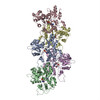
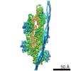
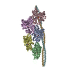
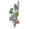
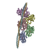
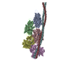
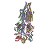
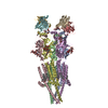
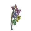
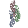
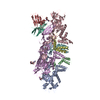
| タイトル | Structure of the F-actin-tropomyosin complex | ||||||
 要素 要素 |
| ||||||
 キーワード キーワード | STRUCTURAL PROTEIN/HYDROLASE / contractile filament / muscle / thin filament / cytoskeleton / structural protein-hydrolase complex | ||||||
| 機能・相同性 |  機能・相同性情報 機能・相同性情報cytoskeletal motor activator activity / myosin heavy chain binding / tropomyosin binding / actin filament bundle / troponin I binding / filamentous actin / mesenchyme migration / skeletal muscle myofibril / actin filament bundle assembly / striated muscle thin filament ...cytoskeletal motor activator activity / myosin heavy chain binding / tropomyosin binding / actin filament bundle / troponin I binding / filamentous actin / mesenchyme migration / skeletal muscle myofibril / actin filament bundle assembly / striated muscle thin filament / skeletal muscle thin filament assembly / actin monomer binding / skeletal muscle fiber development / stress fiber / titin binding / actin filament polymerization / filopodium / actin filament / 加水分解酵素; 酸無水物に作用; 酸無水物に作用・細胞または細胞小器官の運動に関与 / calcium-dependent protein binding / lamellipodium / cell body / protein domain specific binding / hydrolase activity / calcium ion binding / positive regulation of gene expression / magnesium ion binding / ATP binding / identical protein binding / cytoplasm 類似検索 - 分子機能 | ||||||
| 生物種 |  | ||||||
| 手法 | 電子顕微鏡法 / らせん対称体再構成法 / クライオ電子顕微鏡法 / 解像度: 3.7 Å | ||||||
 データ登録者 データ登録者 | von der Ecken, J. / Mueller, M. / Lehman, W. / Manstein, J.M. / Penczek, A.P. / Raunser, S. | ||||||
 引用 引用 | ジャーナル: Nature / 年: 2015 タイトル: Structure of the F-actin-tropomyosin complex. 著者: Julian von der Ecken / Mirco Müller / William Lehman / Dietmar J Manstein / Pawel A Penczek / Stefan Raunser /   要旨: Filamentous actin (F-actin) is the major protein of muscle thin filaments, and actin microfilaments are the main component of the eukaryotic cytoskeleton. Mutations in different actin isoforms lead ...Filamentous actin (F-actin) is the major protein of muscle thin filaments, and actin microfilaments are the main component of the eukaryotic cytoskeleton. Mutations in different actin isoforms lead to early-onset autosomal dominant non-syndromic hearing loss, familial thoracic aortic aneurysms and dissections, and multiple variations of myopathies. In striated muscle fibres, the binding of myosin motors to actin filaments is mainly regulated by tropomyosin and troponin. Tropomyosin also binds to F-actin in smooth muscle and in non-muscle cells and stabilizes and regulates the filaments there in the absence of troponin. Although crystal structures for monomeric actin (G-actin) are available, a high-resolution structure of F-actin is still missing, hampering our understanding of how disease-causing mutations affect the function of thin muscle filaments and microfilaments. Here we report the three-dimensional structure of F-actin at a resolution of 3.7 Å in complex with tropomyosin at a resolution of 6.5 Å, determined by electron cryomicroscopy. The structure reveals that the D-loop is ordered and acts as a central region for hydrophobic and electrostatic interactions that stabilize the F-actin filament. We clearly identify map density corresponding to ADP and Mg(2+) and explain the possible effect of prominent disease-causing mutants. A comparison of F-actin with G-actin reveals the conformational changes during filament formation and identifies the D-loop as their key mediator. We also confirm that negatively charged tropomyosin interacts with a positively charged groove on F-actin. Comparison of the position of tropomyosin in F-actin-tropomyosin with its position in our previously determined F-actin-tropomyosin-myosin structure reveals a myosin-induced transition of tropomyosin. Our results allow us to understand the role of individual mutations in the genesis of actin- and tropomyosin-related diseases and will serve as a strong foundation for the targeted development of drugs. | ||||||
| 履歴 |
| ||||||
| Remark 700 | SHEET DETERMINATION METHOD: AUTHOR DETERMINED |
- 構造の表示
構造の表示
| ムービー |
ムービービューア |
|---|---|
| 構造ビューア | 分子: MolmilJmol/JSmol |
- ダウンロードとリンク
-ダウンロード
| PDBx/mmCIF形式 | 3j8a.cif.gz | 391.3 KB | 表示 | PDBx/mmCIF形式 |
|---|---|---|---|---|
| PDB形式 | pdb3j8a.ent.gz | 315.2 KB | 表示 | PDB形式 |
| PDBx/mmJSON形式 | 3j8a.json.gz | ツリー表示 | PDBx/mmJSON形式 | |
| その他 |  その他のダウンロード その他のダウンロード |
-検証レポート
| アーカイブディレクトリ | https://data.pdbj.org/pub/pdb/validation_reports/j8/3j8aftp://data.pdbj.org/pub/pdb/validation_reports/j8/3j8a | HTTPS FTP |
|---|
-関連構造データ
-リンク
 PDBj
PDBj


- 集合体
集合体
| 登録構造単位 | 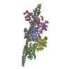
| ||||||||||||||||||||||||||||||||||||||||||
|---|---|---|---|---|---|---|---|---|---|---|---|---|---|---|---|---|---|---|---|---|---|---|---|---|---|---|---|---|---|---|---|---|---|---|---|---|---|---|---|---|---|---|---|
| 1 |
| ||||||||||||||||||||||||||||||||||||||||||
| 対称性 | らせん対称: (回転対称性: 1 / Dyad axis: no / N subunits divisor: 1 / Num. of operations: 1 / Rise per n subunits: 27.5 Å / Rotation per n subunits: -166.4 °) | ||||||||||||||||||||||||||||||||||||||||||
| 非結晶学的対称性 (NCS) | NCSドメイン:
NCSドメイン領域: Component-ID: 1 / Ens-ID: 1 / Beg auth comp-ID: THR / Beg label comp-ID: THR / End auth comp-ID: HIS / End label comp-ID: HIS / Auth seq-ID: 5 - 371 / Label seq-ID: 5 - 371
| ||||||||||||||||||||||||||||||||||||||||||
| 詳細 | The full filament can be generated using the provided helical parameters from a small repeating subunit comprising chain A and residues 145-184 of chains F and G. |
-要素
| #1: タンパク質 | 分子量: 11507.176 Da / 分子数: 2 / 断片: SEE REMARK 999 / 由来タイプ: 組換発現 / 由来: (組換発現)  キーワード: SEE REMARK 999 キーワード: SEE REMARK 999#2: タンパク質 | 分子量: 41875.633 Da / 分子数: 5 / 由来タイプ: 天然 / 由来: (天然) #3: 化合物 | ChemComp-MG /   分子量: 24.305 Da / 分子数: 5 / 由来タイプ: 合成 / 式: Mg 分子量: 24.305 Da / 分子数: 5 / 由来タイプ: 合成 / 式: Mg#4: 化合物 | ChemComp-ADP /   分子量: 427.201 Da / 分子数: 5 / 由来タイプ: 合成 / 式: C10H15N5O10P2 / コメント: ADP, エネルギー貯蔵分子*YM 分子量: 427.201 Da / 分子数: 5 / 由来タイプ: 合成 / 式: C10H15N5O10P2 / コメント: ADP, エネルギー貯蔵分子*YMHas protein modification | Y | 配列の詳細 | MOUSE TROPOMYSIN WAS USED (UNP P58771, RESIDUES 97-231). DUE TO THE LIMITED RESOLUTION OF THE CRYO- ...MOUSE TROPOMYSIN | |
|---|
-実験情報
-実験
| 実験 | 手法: 電子顕微鏡法 |
|---|---|
| EM実験 | 試料の集合状態: FILAMENT / 3次元再構成法: らせん対称体再構成法 |
- 試料調製
試料調製
| 構成要素 |
| ||||||||||||||||||||
|---|---|---|---|---|---|---|---|---|---|---|---|---|---|---|---|---|---|---|---|---|---|
| 緩衝液 | 名称: 5 mM Tris-HCl, pH 7.5, 1 mM DTT, 100 mM KCl, 2 mM MgCl2 pH: 7.5 詳細: 5 mM Tris-HCl, pH 7.5, 1 mM DTT, 100 mM KCl, 2 mM MgCl2 | ||||||||||||||||||||
| 試料 | 包埋: NO / シャドウイング: NO / 染色: NO / 凍結: YES | ||||||||||||||||||||
| 試料支持 | 詳細: C-flats 2/1 copper 300 mesh, Protochips, glow-discharged | ||||||||||||||||||||
| 急速凍結 | 装置: GATAN CRYOPLUNGE 3 / 凍結剤: ETHANE / Temp: 106 K / 湿度: 90 % 詳細: Sample was applied to grid, incubated for 10 seconds, and manually blotted for 3 seconds from the backside with filter paper before plunging into liquid ethane (GATAN CRYOPLUNGE 3) 手法: Sample was applied to grid, incubated for 10 seconds, and manually blotted for 3 seconds from the backside with filter paper. |
- 電子顕微鏡撮影
| 実験機器 |  モデル: Titan Krios / 画像提供: FEI Company |
|---|---|
| 顕微鏡 | モデル: FEI TITAN KRIOS / 日付: 2013年10月17日 / 詳細: Cs-corrected microscope |
| 電子銃 | 電子線源:  FIELD EMISSION GUN / 加速電圧: 300 kV / 照射モード: OTHER FIELD EMISSION GUN / 加速電圧: 300 kV / 照射モード: OTHER |
| 電子レンズ | モード: BRIGHT FIELD / 倍率(公称値): 59000 X / 最大 デフォーカス(公称値): 2600 nm / 最小 デフォーカス(公称値): 800 nm / Cs: 0 mm / カメラ長: 0 mm |
| 試料ホルダ | 試料ホルダーモデル: FEI TITAN KRIOS AUTOGRID HOLDER 傾斜角・最大: 0 ° / 傾斜角・最小: 0 ° |
| 撮影 | 電子線照射量: 14.6 e/Å2 フィルム・検出器のモデル: FEI FALCON II (4k x 4k) |
| 画像スキャン | デジタル画像の数: 1311 |
| 放射 | プロトコル: SINGLE WAVELENGTH / 単色(M)・ラウエ(L): M / 散乱光タイプ: x-ray |
| 放射波長 | 相対比: 1 |
- 解析
解析
| EMソフトウェア |
| ||||||||||||||||||||||||||||||||||||||||||
|---|---|---|---|---|---|---|---|---|---|---|---|---|---|---|---|---|---|---|---|---|---|---|---|---|---|---|---|---|---|---|---|---|---|---|---|---|---|---|---|---|---|---|---|
| CTF補正 | 詳細: each micrograph | ||||||||||||||||||||||||||||||||||||||||||
| らせん対称 | 回転角度/サブユニット: 166.4 ° / 軸方向距離/サブユニット: 27.5 Å / らせん対称軸の対称性: C1 / 詳細: none | ||||||||||||||||||||||||||||||||||||||||||
| 3次元再構成 | 手法: helicon, cter / 解像度: 3.7 Å / 解像度の算出法: FSC 0.5 CUT-OFF / ピクセルサイズ(公称値): 1.14 Å / ピクセルサイズ(実測値): 1.12 Å 詳細: The tropomyosin map filtered to 6.5 Angstrom was merged with the final F-actin map (3.7 Angstrom) to obtain a map of the entire F-actin tropomyosin complex. Coordinates must be shifted by (0. ...詳細: The tropomyosin map filtered to 6.5 Angstrom was merged with the final F-actin map (3.7 Angstrom) to obtain a map of the entire F-actin tropomyosin complex. Coordinates must be shifted by (0.123 -0.381 0.273) to match coordinates to fibre diffraction standards (filament axis = z axis passing through x=0 and y=0). 対称性のタイプ: HELICAL | ||||||||||||||||||||||||||||||||||||||||||
| 原子モデル構築 |
| ||||||||||||||||||||||||||||||||||||||||||
| 原子モデル構築 | 3D fitting-ID: 1 / Accession code: 4A7N / Initial refinement model-ID: 1 / PDB-ID: 4A7N / Source name: PDB / タイプ: experimental model
| ||||||||||||||||||||||||||||||||||||||||||
| 精密化 | 解像度: 3.7→95.2 Å / SU ML: 0.2 / σ(F): 100 / 位相誤差: 26.02 / 立体化学のターゲット値: MLHL
| ||||||||||||||||||||||||||||||||||||||||||
| 溶媒の処理 | 減衰半径: 0.9 Å / VDWプローブ半径: 1.11 Å / 溶媒モデル: FLAT BULK SOLVENT MODEL | ||||||||||||||||||||||||||||||||||||||||||
| 原子変位パラメータ | Biso max: 129.39 Å2 / Biso mean: 50.7811 Å2 / Biso min: 0 Å2 | ||||||||||||||||||||||||||||||||||||||||||
| 精密化ステップ | サイクル: LAST / 解像度: 3.7→95.2 Å
| ||||||||||||||||||||||||||||||||||||||||||
| 拘束条件 |
| ||||||||||||||||||||||||||||||||||||||||||
| Refine LS restraints NCS |
| ||||||||||||||||||||||||||||||||||||||||||
| LS精密化 シェル | 解像度: 3.7→95.2 Å / Total num. of bins used: 1
|