 Movie
Movie Controller


+ Open data

- Basic information
| Entry | Database: PDB / ID: 3j6b | |||||||||
|---|---|---|---|---|---|---|---|---|---|---|
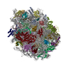
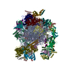
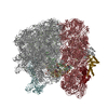
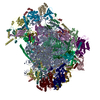
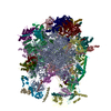
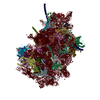
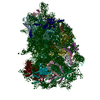
| Title | Structure of the yeast mitochondrial large ribosomal subunit | |||||||||
 Components Components |
| |||||||||
 Keywords Keywords | RIBOSOME / mitochondrial ribosome / large subunit / protein-RNA complex | |||||||||
| Function / homology |  Function and homology information Function and homology informationpositive regulation of mitochondrial DNA replication / mitochondrial cytochrome c oxidase assembly / Mitochondrial protein degradation / DNA strand exchange activity / mitochondrial genome maintenance / ribonuclease III activity / mitochondrial large ribosomal subunit / mitochondrial translation / RNA processing / cell redox homeostasis ...positive regulation of mitochondrial DNA replication / mitochondrial cytochrome c oxidase assembly / Mitochondrial protein degradation / DNA strand exchange activity / mitochondrial genome maintenance / ribonuclease III activity / mitochondrial large ribosomal subunit / mitochondrial translation / RNA processing / cell redox homeostasis / ribosome biogenesis / large ribosomal subunit / single-stranded DNA binding / double-stranded RNA binding / transferase activity / cellular response to oxidative stress / large ribosomal subunit rRNA binding / DNA recombination / cytosolic large ribosomal subunit / mitochondrial inner membrane / negative regulation of translation / rRNA binding / ribosome / structural constituent of ribosome / translation / mRNA binding / regulation of DNA-templated transcription / mitochondrion / DNA binding / RNA binding / zinc ion binding / nucleus / cytoplasm Similarity search - Function | |||||||||
| Biological species |  | |||||||||
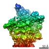
| Method | ELECTRON MICROSCOPY / single particle reconstruction / cryo EM / Resolution: 3.2 Å | |||||||||
 Authors Authors | Amunts, A. / Brown, A. / Bai, X.C. / Llacer, J.L. / Hussain, T. / Emsley, P. / Long, F. / Murshudov, G. / Scheres, S.H.W. / Ramakrishnan, V. | |||||||||
 Citation Citation | Journal: Science / Year: 2014 Title: Structure of the yeast mitochondrial large ribosomal subunit. Authors: Alexey Amunts / Alan Brown / Xiao-Chen Bai / Jose L Llácer / Tanweer Hussain / Paul Emsley / Fei Long / Garib Murshudov / Sjors H W Scheres / V Ramakrishnan /  Abstract: Mitochondria have specialized ribosomes that have diverged from their bacterial and cytoplasmic counterparts. We have solved the structure of the yeast mitoribosomal large subunit using single- ...Mitochondria have specialized ribosomes that have diverged from their bacterial and cytoplasmic counterparts. We have solved the structure of the yeast mitoribosomal large subunit using single-particle cryo-electron microscopy. The resolution of 3.2 angstroms enabled a nearly complete atomic model to be built de novo and refined, including 39 proteins, 13 of which are unique to mitochondria, as well as expansion segments of mitoribosomal RNA. The structure reveals a new exit tunnel path and architecture, unique elements of the E site, and a putative membrane docking site. | |||||||||
| History |
|
- Structure visualization
| Movie |
Movie viewer |
|---|---|
| Structure viewer | Molecule: MolmilJmol/JSmol |
- Downloads & links
-Download
| PDBx/mmCIF format | 3j6b.cif.gz | 4.6 MB | Display | PDBx/mmCIF format |
|---|---|---|---|---|
| PDB format | pdb3j6b.ent.gz | Display | PDB format | |
| PDBx/mmJSON format | 3j6b.json.gz | Tree view | PDBx/mmJSON format | |
| Others |  Other downloads Other downloads |
-Validation report
| Summary document | 3j6b_validation.pdf.gz | 1.1 MB | Display | wwPDB validaton report |
|---|---|---|---|---|
| Full document | 3j6b_full_validation.pdf.gz | 1.3 MB | Display | |
| Data in XML | 3j6b_validation.xml.gz | 202.8 KB | Display | |
| Data in CIF | 3j6b_validation.cif.gz | 346.9 KB | Display | |
| Arichive directory | https://data.pdbj.org/pub/pdb/validation_reports/j6/3j6bftp://data.pdbj.org/pub/pdb/validation_reports/j6/3j6b | HTTPS FTP |
-Related structure data
| Related structure data | 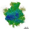 2566MC M: map data used to model this data C: citing same article ( |
|---|---|
| Similar structure data |
-Links
 PDBj
PDBj


- Assembly
| Deposited unit | 
|
|---|---|
| 1 |
|
-Components
-RNA chain , 2 types, 2 molecules Ae
| #1: RNA chain | Mass: 1057805.375 Da / Num. of mol.: 1 / Source method: isolated from a natural source / Source: (natural) |
|---|---|
| #41: RNA chain | Mass: 6451.927 Da / Num. of mol.: 1 / Source method: isolated from a natural source / Source: (natural) |
+54S ribosomal protein ... , 37 types, 37 molecules BCDEFGHIJKLMNOPQRSTUVWXY012345...
-Protein , 2 types, 2 molecules Zd
| #26: Protein | Mass: 13276.667 Da / Num. of mol.: 1 / Source method: isolated from a natural source / Source: (natural) |
|---|---|
| #40: Protein | Mass: 26937.762 Da / Num. of mol.: 1 / Source method: isolated from a natural source / Source: (natural) |
-Non-polymers , 3 types, 113 molecules 


| #42: Chemical | ChemComp-MG /  Mass: 24.305 Da / Num. of mol.: 110 / Source method: obtained synthetically / Formula: Mg Mass: 24.305 Da / Num. of mol.: 110 / Source method: obtained synthetically / Formula: Mg#43: Chemical | ChemComp-NA / |  Mass: 22.990 Da / Num. of mol.: 1 / Source method: obtained synthetically / Formula: Na Mass: 22.990 Da / Num. of mol.: 1 / Source method: obtained synthetically / Formula: Na#44: Chemical |  Mass: 65.409 Da / Num. of mol.: 2 / Source method: obtained synthetically / Formula: Zn Mass: 65.409 Da / Num. of mol.: 2 / Source method: obtained synthetically / Formula: Zn |
|---|
-Experimental details
-Experiment
| Experiment | Method: ELECTRON MICROSCOPY |
|---|---|
| EM experiment | Aggregation state: PARTICLE / 3D reconstruction method: single particle reconstruction |
- Sample preparation
| Component | Name: Yeast Mitochondrial Large Ribosomal Subunit / Type: RIBOSOME |
|---|---|
| Molecular weight | Value: 1.9 MDa / Experimental value: NO |
| Buffer solution | Name: 20 mM HEPES-KOH, pH 7.5, 100 mM potassium chloride, 20 mM magnesium acetate, 2 mM DTT pH: 7.5 Details: 20 mM HEPES-KOH, pH 7.5, 100 mM potassium chloride, 20 mM magnesium acetate, 2 mM DTT |
| Specimen | Conc.: 0.15 mg/ml / Embedding applied: NO / Shadowing applied: NO / Staining applied: NO / Vitrification applied: YES |
| Specimen support | Details: glow-discharged holey carbon grids (Quantifoil R2/2) with home-made continuous carbon film |
| Vitrification | Instrument: FEI VITROBOT MARK II / Cryogen name: ETHANE / Temp: 90 K / Humidity: 100 % Details: Blot 2.5 seconds before plunging into liquid ethane (FEI VITROBOT MARK II) Method: Blot 2.5 seconds before plunging |
- Electron microscopy imaging
| Experimental equipment |  Model: Titan Krios / Image courtesy: FEI Company |
|---|---|
| Microscopy | Model: FEI TITAN KRIOS / Date: May 25, 2013 Details: Objective lens astigmatism was corrected at 59,000 times magnification |
| Electron gun | Electron source:  FIELD EMISSION GUN / Accelerating voltage: 300 kV / Illumination mode: FLOOD BEAM FIELD EMISSION GUN / Accelerating voltage: 300 kV / Illumination mode: FLOOD BEAM |
| Electron lens | Mode: BRIGHT FIELD / Nominal magnification: 59000 X / Nominal defocus max: 4700 nm / Nominal defocus min: 1200 nm |
| Specimen holder | Specimen holder model: FEI TITAN KRIOS AUTOGRID HOLDER / Specimen holder type: FEI TITAN KRIOS AUTOGRID HOLDER / Temperature: 85 K / Temperature (max): 90 K |
| Image recording | Electron dose: 25 e/Å2 / Film or detector model: FEI FALCON II (4k x 4k) |
| Image scans | Num. digital images: 1030 |
| Radiation wavelength | Relative weight: 1 |
- Processing
| Software | Name: REFMAC / Version: 5.8.0067 2014/01/21 / Classification: refinement / Contact author: Garib N. Murshudov / Contact author email: garib[at]mrc-lmb.cam.ac.uk Description: (un)restrained refinement or idealisation of macromolecular structures | |||||||||||||||||||||||||||||||||||||||||||||||||||||||||||||||||||||||||||||||||||||||||||||||||||||||||
|---|---|---|---|---|---|---|---|---|---|---|---|---|---|---|---|---|---|---|---|---|---|---|---|---|---|---|---|---|---|---|---|---|---|---|---|---|---|---|---|---|---|---|---|---|---|---|---|---|---|---|---|---|---|---|---|---|---|---|---|---|---|---|---|---|---|---|---|---|---|---|---|---|---|---|---|---|---|---|---|---|---|---|---|---|---|---|---|---|---|---|---|---|---|---|---|---|---|---|---|---|---|---|---|---|---|---|
| EM software |
| |||||||||||||||||||||||||||||||||||||||||||||||||||||||||||||||||||||||||||||||||||||||||||||||||||||||||
| CTF correction | Details: Each particle | |||||||||||||||||||||||||||||||||||||||||||||||||||||||||||||||||||||||||||||||||||||||||||||||||||||||||
| Symmetry | Point symmetry: C1 (asymmetric) | |||||||||||||||||||||||||||||||||||||||||||||||||||||||||||||||||||||||||||||||||||||||||||||||||||||||||
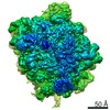
| 3D reconstruction | Resolution: 3.2 Å / Resolution method: FSC 0.143 CUT-OFF / Num. of particles: 47124 / Nominal pixel size: 1.34 Å / Actual pixel size: 1.34 Å Details: A newly developed statistical movie processing approach was used to compensate for beam-induced movement. Refinement type: HALF-MAPS REFINED INDEPENDENTLY / Symmetry type: POINT | |||||||||||||||||||||||||||||||||||||||||||||||||||||||||||||||||||||||||||||||||||||||||||||||||||||||||
| Refinement | Resolution: 3.2→262.64 Å / Cor.coef. Fo:Fc: 0.938 / SU B: 16.551 / SU ML: 0.26 / Average fsc overall: 0.8808 / Average fsc work: 0.8808 / ESU R: 0.372 Details: HYDROGENS HAVE BEEN ADDED IN THEIR RIDING POSITIONS
| |||||||||||||||||||||||||||||||||||||||||||||||||||||||||||||||||||||||||||||||||||||||||||||||||||||||||
| Solvent computation | Ion probe radii: 0.8 Å / Shrinkage radii: 0.8 Å / VDW probe radii: 1.2 Å / Solvent model: BABINET MODEL PLUS MASK | |||||||||||||||||||||||||||||||||||||||||||||||||||||||||||||||||||||||||||||||||||||||||||||||||||||||||
| Displacement parameters | Biso mean: 94.596 Å2
| |||||||||||||||||||||||||||||||||||||||||||||||||||||||||||||||||||||||||||||||||||||||||||||||||||||||||
| Refine LS restraints |
|