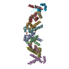
Journal: J Mol Biol / Year: 2012 Title: Filaments from Ignicoccus hospitalis show diversity of packing in proteins containing N-terminal type IV pilin helices. Authors: Xiong Yu / Charles Goforth / Carolin Meyer / Reinhard Rachel / Reinhard Wirth / Gunnar F Schröder / Edward H Egelman / Abstract: Bacterial motility is driven by the rotation of flagellar filaments that supercoil. The supercoiling involves the switching of coiled-coil protofilaments between two different states. In archaea, the ...Bacterial motility is driven by the rotation of flagellar filaments that supercoil. The supercoiling involves the switching of coiled-coil protofilaments between two different states. In archaea, the flagellar filaments responsible for motility are formed by proteins with distinct homology in their N-terminal portion to bacterial Type IV pilins. The bacterial pilins have a single N-terminal hydrophobic α-helix, not the coiled coil found in flagellin. We have used electron cryo-microscopy to study the adhesion filaments from the archaeon Ignicoccus hospitalis. While I. hospitalis is non-motile, these filaments make transitions between rigid stretches and curved regions and appear morphologically similar to true archaeal flagellar filaments. A resolution of ~7.5Å allows us to unambiguously build a model for the packing of these N-terminal α-helices, and this packing is different from several bacterial Type IV pili whose structure has been analyzed by electron microscopy and modeling. Our results show that the mechanism responsible for the supercoiling of bacterial flagellar filaments cannot apply to archaeal filaments.
THE ASSEMBLY REPRESENTED IN THIS ENTRY HAS REGULAR HELICAL SYMMETRY WITH THE FOLLOWING PARAMETERS: ROTATION PER SUBUNIT (TWIST) = 106.65 DEGREES; RISE PER SUBUNIT (HEIGHT) = 5.3 ANGSTROM
-
Components
#1: Protein/peptide
... archaealadhesionfilamentcore
Mass: 2710.340 Da / Num. of mol.: 21 / Fragment: SEE REMARK 999 / Source method: isolated from a natural source / Source: (natural) Ignicoccus hospitalis (archaea) / References: UniProt: A8AAA0
Sequence details
THOUGH THE FULL PROTEIN (UNP RESIDUES 1-310) WAS PRESENT, ONLY RESIDUES 8-33 WERE MODELED.
-
Experimental details
-
Experiment
Experiment
Method: ELECTRON MICROSCOPY
EM experiment
Aggregation state: FILAMENT / 3D reconstruction method: helical reconstruction
-
Sample preparation
Component
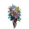
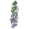
Name: Adhesion filament / Type: COMPLEX
Specimen
Embedding applied: NO / Shadowing applied: NO / Staining applied: NO / Vitrification applied: YES
In the structure databanks used in Yorodumi, some data are registered as the other names, "COVID-19 virus" and "2019-nCoV". Here are the details of the virus and the list of structure data.
Jan 31, 2019. EMDB accession codes are about to change! (news from PDBe EMDB page)
EMDB accession codes are about to change! (news from PDBe EMDB page)
The allocation of 4 digits for EMDB accession codes will soon come to an end. Whilst these codes will remain in use, new EMDB accession codes will include an additional digit and will expand incrementally as the available range of codes is exhausted. The current 4-digit format prefixed with “EMD-” (i.e. EMD-XXXX) will advance to a 5-digit format (i.e. EMD-XXXXX), and so on. It is currently estimated that the 4-digit codes will be depleted around Spring 2019, at which point the 5-digit format will come into force.
The EM Navigator/Yorodumi systems omit the EMD- prefix.
Related info.:Q: What is EMD? / ID/Accession-code notation in Yorodumi/EM Navigator
Yorodumi is a browser for structure data from EMDB, PDB, SASBDB, etc.
This page is also the successor to EM Navigator detail page, and also detail information page/front-end page for Omokage search.
The word "yorodu" (or yorozu) is an old Japanese word meaning "ten thousand". "mi" (miru) is to see.
Related info.:EMDB / PDB / SASBDB / Comparison of 3 databanks / Yorodumi Search / Aug 31, 2016. New EM Navigator & Yorodumi / Yorodumi Papers / Jmol/JSmol / Function and homology information / Changes in new EM Navigator and Yorodumi
 Movie
Movie Controller

 Yorodumi
 Open data
 Basic information
 Components
 Keywords
 Function and homology information
 Ignicoccus hospitalis (archaea)
 Authors
 Citation
 Structure visualization
 Downloads & links
 Other downloads


 PDBj
PDBj Assembly
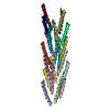
 Sample preparation
 Electron microscopy imaging
 FIELD EMISSION GUN / Accelerating voltage: 200 kV / Illumination mode: FLOOD BEAM
 Processing