 Movie
Movie Controller

[English] 日本語
 Yorodumi
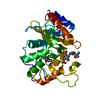
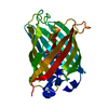
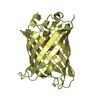
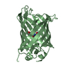
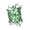
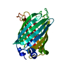
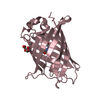
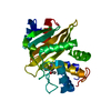
- PDB-3iwc: T. maritima AdoMetDC complex with S-Adenosylmethionine methyl ester -
+ Open data

- Basic information
| Entry | Database: PDB / ID: 3iwc | |||||||||
|---|---|---|---|---|---|---|---|---|---|---|
| Title | T. maritima AdoMetDC complex with S-Adenosylmethionine methyl ester | |||||||||
 Components Components | (S-adenosylmethionine decarboxylase) x 2 | |||||||||
 Keywords Keywords | LYASE / Autocatalytic cleavage / Decarboxylase / Polyamine biosynthesis / Pyruvate / S-adenosyl-L-methionine / Schiff base / Spermidine biosynthesis / Zymogen | |||||||||
| Function / homology |  Function and homology information Function and homology informationadenosylmethionine decarboxylase / adenosylmethionine decarboxylase activity / spermidine biosynthetic process / cytosol Similarity search - Function | |||||||||
| Biological species |   Thermotoga maritima (bacteria) Thermotoga maritima (bacteria) | |||||||||
| Method |  X-RAY DIFFRACTION / SYNCHROTRON / MOLECULAR REPLACEMENT / Resolution: 1.9 Å X-RAY DIFFRACTION / SYNCHROTRON / MOLECULAR REPLACEMENT / Resolution: 1.9 Å | |||||||||
 Authors Authors | Bale, S. / Kavita, B. / Ealick, S.E. | |||||||||
 Citation Citation | Journal: Acta Crystallogr.,Sect.D / Year: 2010 Title: Complexes of Thermotoga maritimaS-adenosylmethionine decarboxylase provide insights into substrate specificity. Authors: Bale, S. / Baba, K. / McCloskey, D.E. / Pegg, A.E. / Ealick, S.E. | |||||||||
| History |
|
- Structure visualization
| Structure viewer | Molecule: MolmilJmol/JSmol |
|---|
- Downloads & links
-Download
| PDBx/mmCIF format | 3iwc.cif.gz | 66 KB | Display | PDBx/mmCIF format |
|---|---|---|---|---|
| PDB format | pdb3iwc.ent.gz | 47.6 KB | Display | PDB format |
| PDBx/mmJSON format | 3iwc.json.gz | Tree view | PDBx/mmJSON format | |
| Others |  Other downloads Other downloads |
-Validation report
| Arichive directory | https://data.pdbj.org/pub/pdb/validation_reports/iw/3iwcftp://data.pdbj.org/pub/pdb/validation_reports/iw/3iwc | HTTPS FTP |
|---|
-Related structure data
| Related structure data |  3iwbC  3iwdC  1tluS S: Starting model for refinement C: citing same article ( |
|---|---|
| Similar structure data |


-Links
 PDBj
PDBj- Assembly
| Deposited unit | 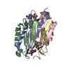
| ||||||||
|---|---|---|---|---|---|---|---|---|---|
| 1 |
| ||||||||
| 2 | 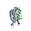
| ||||||||
| 3 | 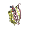
| ||||||||
| Unit cell |
| ||||||||
| Details | biological assembly is a dimer in the asymmetric unit |
-Components
| #1: Protein | Mass: 6999.928 Da / Num. of mol.: 2 / Fragment: residues 1-62 Source method: isolated from a genetically manipulated source Source: (gene. exp.) Thermotoga maritima (bacteria) / Gene: speH, TM_0655 / Plasmid: pTmSpeD.28 / Production host: References: UniProt: Q9WZC3, adenosylmethionine decarboxylase #2: Protein | Mass: 7807.784 Da / Num. of mol.: 2 / Fragment: residues 64-130 Source method: isolated from a genetically manipulated source Source: (gene. exp.) Thermotoga maritima (bacteria) / Gene: speH, TM_0655 / Plasmid: pTmSpeD.28 / Production host: References: UniProt: Q9WZC3, adenosylmethionine decarboxylase #3: Chemical | 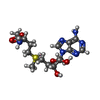  Mass: 414.480 Da / Num. of mol.: 2 / Source method: obtained synthetically / Formula: C16H26N6O5S Mass: 414.480 Da / Num. of mol.: 2 / Source method: obtained synthetically / Formula: C16H26N6O5S#4: Water | ChemComp-HOH / |  Mass: 18.015 Da / Num. of mol.: 108 / Source method: isolated from a natural source / Formula: H2O Mass: 18.015 Da / Num. of mol.: 108 / Source method: isolated from a natural source / Formula: H2OHas protein modification | Y | |
|---|
-Experimental details
-Experiment
| Experiment | Method: X-RAY DIFFRACTION / Number of used crystals: 1 |
|---|
- Sample preparation
| Crystal | Density Matthews: 2.52 Å3/Da / Density % sol: 51.18 % |
|---|---|
| Crystal grow | Temperature: 298 K / Method: vapor diffusion, hanging drop / pH: 8 Details: 2.8 M ammonium formate, 100 mM HEPES, pH 8.0, vapor diffusion, hanging drop, temperature 298K |
-Data collection
| Diffraction | Mean temperature: 100 K |
|---|---|
| Diffraction source | Source: SYNCHROTRON / Site: APS  / Beamline: 24-ID-E / Wavelength: 0.9792 Å / Beamline: 24-ID-E / Wavelength: 0.9792 Å |
| Detector | Type: ADSC QUANTUM 315 / Detector: CCD / Date: Mar 1, 2007 |
| Radiation | Protocol: SINGLE WAVELENGTH / Monochromatic (M) / Laue (L): M / Scattering type: x-ray |
| Radiation wavelength | Wavelength: 0.9792 Å / Relative weight: 1 |
| Reflection | Resolution: 1.9→50 Å / Num. all: 22459 / Num. obs: 22259 / % possible obs: 98.9 % / Observed criterion σ(F): 0 / Observed criterion σ(I): 0 / Redundancy: 4.1 % / Biso Wilson estimate: 19.2 Å2 / Rmerge(I) obs: 0.096 / Rsym value: 0.096 / Χ2: 1.317 / Net I/σ(I): 11.2 |
| Reflection shell | Resolution: 1.9→1.97 Å / Redundancy: 2.3 % / Rmerge(I) obs: 0.216 / Mean I/σ(I) obs: 3.1 / Num. unique all: 2067 / Rsym value: 0.216 / Χ2: 0.544 / % possible all: 90.4 |
- Processing
| Software |
| ||||||||||||||||||||||||||||||||||||
|---|---|---|---|---|---|---|---|---|---|---|---|---|---|---|---|---|---|---|---|---|---|---|---|---|---|---|---|---|---|---|---|---|---|---|---|---|---|
| Refinement | Method to determine structure: MOLECULAR REPLACEMENT Starting model: 1TLU Resolution: 1.9→32.7 Å / Rfactor Rfree error: 0.005 / Occupancy max: 1 / Occupancy min: 1 / Data cutoff high absF: 86489 / Data cutoff low absF: 0 / Isotropic thermal model: RESTRAINED / Cross valid method: THROUGHOUT / σ(F): 0 / σ(I): 0 / Stereochemistry target values: Engh & Huber / Details: BULK SOLVENT MODEL USED
| ||||||||||||||||||||||||||||||||||||
| Solvent computation | Solvent model: FLAT MODEL / Bsol: 60.519 Å2 / ksol: 0.4 e/Å3 | ||||||||||||||||||||||||||||||||||||
| Displacement parameters | Biso max: 80.4 Å2 / Biso mean: 32.829 Å2 / Biso min: 14.07 Å2
| ||||||||||||||||||||||||||||||||||||
| Refine analyze |
| ||||||||||||||||||||||||||||||||||||
| Refinement step | Cycle: LAST / Resolution: 1.9→32.7 Å
| ||||||||||||||||||||||||||||||||||||
| Refine LS restraints |
| ||||||||||||||||||||||||||||||||||||
| LS refinement shell | Resolution: 1.9→2.02 Å / Rfactor Rfree error: 0.016 / Total num. of bins used: 6
| ||||||||||||||||||||||||||||||||||||
| Xplor file |
|
